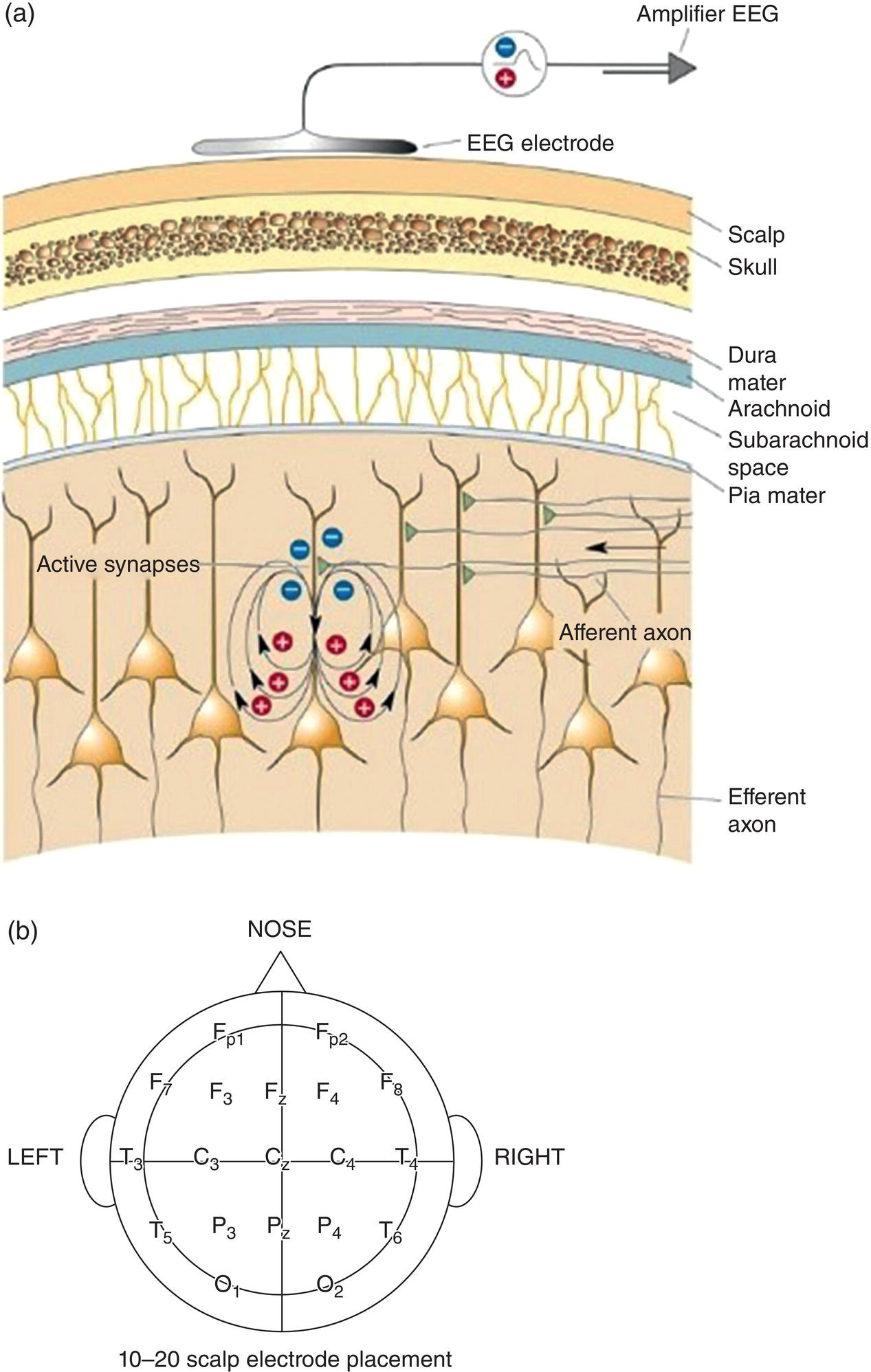
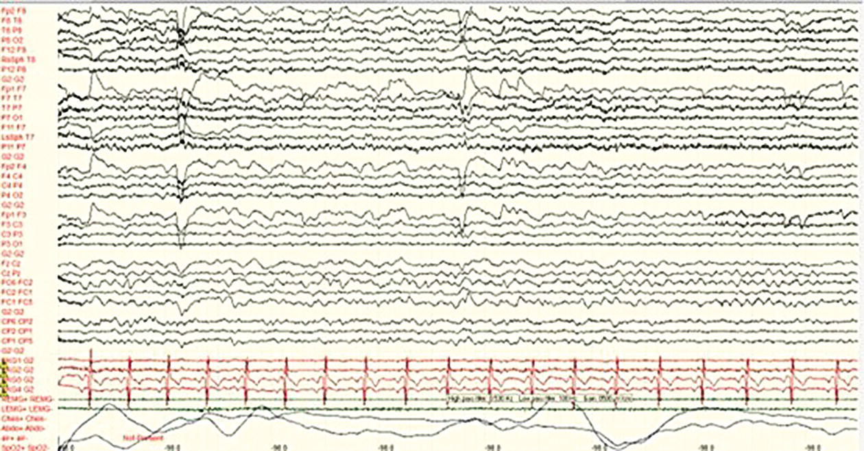
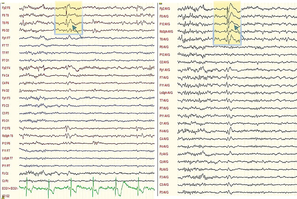
Fahmida A. Chowdhury In 1929, Hans Berger, a German psychiatrist, recorded the first human electroencephalogram (EEG). In the 1940s, Gibbs and colleagues demonstrated 3 Hz spike–wave discharge in ‘petit mal’ epilepsy (now ‘absence’ epilepsy). The EEG is crucial for diagnosing, classifying and managing people with seizures and other neurological disorders. Currently, the majority of EEG reporting is based on visual reading by skilled clinical neurophysiologists. AI‐driven EEG reporting is becoming available and is expected to have a significant impact on EEG reporting. Electrical activity recorded by scalp electrodes reflects the summation of excitatory and inhibitory postsynaptic potentials in apical dendrites of pyramidal neurons in superficial cortex layers (Figure 7.1a). Most of the signal is produced by vertically oriented neurons.1 The EEG signal is measured as the potential difference between two electrodes. It is attenuated, filtered, and potentially contaminated by structures between the cortex and the recording electrode, including CSF, dura, bone, muscle, blood vessels, and skin. Six to ten cm2 of cortex needs to be activated synchronously to generate enough potential for changes to be recorded from the scalp.2 The electrical signal needs to be amplified by a factor of 100,000, and the recording is prone to physiological artefacts (such as ECG, eye blinks, chewing, or other muscle activity) and non‐physiological artefacts (electrical interference). Scalp EEG has high temporal resolution but low spatial resolution, as significant amounts of the cortex, particularly in basal and mesial areas of the hemispheres, are not covered by standard electrode placements. A standard routine EEG recording takes about an hour. Electrodes are applied using the international 10–20 system, which uses electrodes 10% or 20% of the distance between standard landmarks (Figure 7.1b). Additional electrodes may be placed, depending on specific questions, such as surface electromyographic (EMG) electrodes that enable the correlation of muscle activity with EEG activity. ECG electrodes and respiratory belts are used to examine autonomic function and to detect cardiac arrhythmias, which are a crucial part of differential diagnosis in individuals presenting with blackouts (Figure 7.2). The EEG can be displayed using different montages, such as bipolar, in which each waveform in the EEG represents the difference in voltage between two adjacent electrodes and a reference showing the voltage between a specific active electrode and a designated reference (Figure 7.3). FIGURE 7.1 (a) Source of EEG signal. Adapted from Neuroscience: Exploring the brain (4th Edition, 2016). (b) 10–20 scalp electrode placement. F = frontal, P = parietal, T = temporal, O = occipital, C = central, odd numbers indicate left hemisphere and even numbers right hemisphere. FIGURE 7.2 Polygraphic recording showing EEG (bipolar montage) in black with additional frontocentral and centroparietal EEG electrodes (FC1, FC2, CP1, CP2, FC5, FC6, CP5, CP6), and ECG electrodes in red, EMG electrodes in green and respiratory band recording in blue. FIGURE 7.3 Right temporal sharp wave with after‐going slow activity, shown in bipolar (left) and referential montage (right), with ‘phase reversal’ at F8 on bipolar montage and maximal amplitude at F8 on referential montage, indicating this is where the activity is maximal. The standard scalp EEG picks up activity between 1 to 30 Hz (delta 1‐4 Hz, theta 4‐7 Hz, alpha 8–12 Hz, beta 13‐30 Hz) (Table 7.1). Faster frequencies known as high frequency oscillations (HFOs) (gamma 30‐80 Hz, ripples 80‐ 250 Hz, and fast ripples 250‐500 Hz) can be picked up with intracranial EEG, which is not attenuated by scalp and skull. These may be a biomarker for epileptogenic tissues, though physiological HFOs have also been noted in people without epilepsy. Higher‐density scalp EEG, when combined with computational analysis, may provide additional information on the source localisation of activity in focal epilepsy. Table 7.1 Frequency bandwidths on scalp EEG. Hyperventilation and photic stimulation increase the yield of abnormal findings and are included in routine EEG following standardised protocols. These procedures are generally safe; however, there are specific contraindications to consider. Hyperventilation and photic stimulation are associated with a small risk of triggering seizures. The National Institute for Health and Care Excellence (NICE) guidelines recommend hyperventilation for a minimum of 3 minutes, if not contraindicated. Hyperventilation may trigger a seizure in a small number of people, so the individual should provide informed consent before undergoing this procedure.3 It may not be advisable to use hyperventilation in seizure‐free individuals. It should also be avoided in those with significant pulmonary and cardiac disease or raised intracranial pressure. It can be instrumental in the diagnosis of absence epilepsies, as well as for triggering non‐epileptic events. During intermittent photic stimulation, the individual is presented with a series of trains of flashing lights. The photoparoxysmal response occurs when photic stimulation produces bilaterally synchronous epileptiform discharges that outlast the stimulus. The middle flash frequencies, between 10 and 20 Hz, are most provocative. This is most commonly seen in genetic generalised epilepsy but can be seen in some focal epilepsies, and may also exist as a genetic trait in some people without epilepsy.4 There are a range of scalp EEG recordings, each type with its advantages and disadvantages (Table 7.2). Shorter recordings tend to capture interictal abnormalities, whereas longer recordings, ideally with video, are more likely to yield diagnostic information when events need to be captured. Table 7.2 Different types of EEG: advantages and disadvantages. Abnormal findings can be broadly categorised as specific or nonspecific. There are also normal variants of no clinical significance. The EEG of healthy individuals can exhibit a range of spiky, sharpened, or rhythmic features resembling epileptiform activity, particularly during sleep. These are normal variants of no clinical significance and include benign epileptiform sleep transients, lambda waves, Mu rhythm, wicket spikes, 14 and 6 Hz positive bursts, rhythmic mid‐temporal theta, midline theta, and subclinical rhythmic epileptiform discharge in adults (SREDA). These variants are a potential source of confusion, and it is essential to be aware of them to avoid EEG overinterpretation, which can lead to misdiagnosis.5 Slowing of background rhythms is a nonspecific abnormality indicating cortical dysfunction of nonspecific aetiology. A wide range of pathologies, including tumours, stroke, encephalitis, trauma and haematomas, as well as any other structural lesions, may cause them. These are not associated with seizures, though they may also be seen in people with epilepsy, focally or in a generalised distribution. As focal interictal slowing can reflect underlying structural pathology, the presence of this finding should prompt brain imaging. Triphasic waves are non‐epileptiform activity indicative of nonspecific cortical dysfunction, commonly seen in metabolic encephalopathies. Epileptiform activity, which is highly specific for epilepsy, includes spikes, polyspikes, and sharp waves. Their morphology and duration define these: spikes (<70 ms) and sharp waves (70‐200 ms), and reflect the hyperexcitability of a cortical area (Figure 7.3). The criteria for identifying spikes require several features to be present.6 Appear dissimilar to the background waveforms. Routine EEG recordings are unlikely to capture events, and seizures are usually only captured during longer recordings. The exception is absence seizures, which can be triggered by hyperventilation. Ictal patterns are identified by the abrupt onset of epileptiform activity that evolves in frequency, amplitude, and /or spatial distribution and terminates abruptly. Some seizure types have characteristic patterns, such as the 3 Hz spike and wave in absence seizures, the evolving 5–7 Hz temporal theta rhythm in mesial temporal epilepsies, and high‐frequency discharges in tonic and neocortical seizures. Ictal changes may be obscured by artefacts from movement or muscle, particularly in frontal lobe epilepsies with motor features, and also due to the distance from the recording electrodes on the scalp. Scalp EEG commonly shows no change in focal preserved consciousness seizures (auras), because the ictal discharge involves too small a neuronal aggregate for the synchronised activity to be detected on the scalp. The diagnosis of epilepsy is clinical, and EEG has a high false‐negative rate and should not be used to ‘exclude’ epilepsy. EEG also has a low but critical false positive rate, so an abnormal EEG with epileptiform discharges does not diagnose a seizure disorder. Still, EEG is a core investigation in epilepsy and, when used in the correct context, can aid diagnosis and management in many ways. It can be helpful in: The presence of interictal epileptiform discharges (IED) can support a diagnosis of epilepsy in the correct clinical context. Several factors influence the EEG diagnostic yield, including the frequency, severity, and type of epilepsy, as well as the timing of the recording, particularly in generalised epilepsies. IED are more commonly seen in individuals with generalised than focal epilepsies. The site of epileptic focus can affect the result; for example, most people with TLE show IED, whereas epileptic foci in mesial or basal cortical regions remote from scalp electrodes are less likely to. Age is another factor; children are more likely to show epileptiform discharges than older people. Hyperventilation and photic stimulation can increase yield. The diagnostic yield of the EEG can also be increased by sleep deprivation, recording during sleep, serial recordings, or early post‐seizure recording. Temporal sampling is limited, particularly in shorter‐duration recordings, such as routine EEG. The sensitivity of a single routine EEG is 50%, and if a sleep EEG follows, this can be increased to 80%. With repeated recordings, the sensitivity rises to 90%.7,8 Prolonged sampling using ambulatory EEG monitoring can increase the yield by about 20%.9 The timing of the EEG to the last seizure may also be relevant. An interictal EEG within 24 hours of an episode showed an abnormality in one half, compared with about a third who had a later EEG.10 Caution is necessary when assessing the significance of IED, especially if the clinical history is not suggestive of epilepsy. In aircrew with no known history of seizures, the incidence of IED in a routine EEG was 0.5%.11 This is higher in hospitalised individuals, 2% in a cohort of various pathologies and significantly higher in people with psychiatric diseases at 5%, and 13% in those taking clozapine.12 Table 7.3 Indications for EEG. NICE guidelines recommend routine EEG recording if the clinical history of an event suggests an epileptic seizure. EEG should not be used if the presentation suggests syncope or a non‐epileptic attack (Table 7.3). The test should be performed within four weeks of the request. Following a single unprovoked seizure, if an EEG shows a definite epileptiform discharge, there is a greater risk of seizure recurrence. A systematic review showed that the recurrence risk at two years was about a quarter if the EEG was normal, but almost two‐thirds if epileptiform activity was present.13 The ILAE diagnostic criteria for epilepsy include a single unprovoked seizure with the probability of recurrent seizures of 60% or more. Therefore, an EEG containing an IED following a single seizure in the current clinical setting may confer a diagnosis of epilepsy.14 In focal epilepsy, IED do not provide a reliable severity or seizure frequency index. Reduction in IED only has a weak association with reduced seizure frequency. Hence, EEG is not recommended for assessing treatment effects. The exception is in generalised epilepsy, in which persistent discharges may indicate sub‐optimal therapy, but it is not recommended to ‘treat the EEG’, attempting to abolish all spikes. The role of EEG is uncertain in individuals in remission wishing to withdraw or reduce ASM. The syndrome is the most critical variable in predicting seizure recurrence after ASM withdrawal. Spike–wave discharge in people with generalised epilepsy and evidence of a generalised photo paroxysmal response is associated with a high recurrence risk. The prognostic significance of other patterns and abnormalities in other epilepsy types is less clear.15 Confusional states may result from encephalopathy or non‐convulsive status, and EEG can help differentiate between these. In severe encephalopathy, however, this can be challenging due to the overlap between interictal and ictal patterns.
CHAPTER 7
Neurophysiology
ELECTROENCEPHALOGRAPHY
BASIC PRINCIPLES
EEG FREQUENCY
Rhythm
Frequency (Hz)
Features
Delta
1–4
Sleep
Theta
4–7
Children, adults, drowsiness, sleep
Alpha
8–12
Adult, rest, posterior dominant, responsive to eye closure
Beta
13–10
Adult, frontocentral, mental activation
ACTIVATION TECHNIQUES
EEG IN CLINICAL PRACTICE
TYPES OF SCALP EEG RECORDINGS
EEG type
Advantages
Limitations
Routine
Twenty minutes, with video. Hyperventilation and photic stimulation
Optimal recording conditions, controlled by a physiologist
Low temporal sampling due to the short length of the recording
Sleep/
Sleep deprived
One hour with attempted sleep, with video
Optimal recording conditions, controlled by a physiologist
As above
Ambulatory
24–72 hours, no video
Longer temporal sampling
No video to document clinical features, prone to artefacts
Video‐telemetry
Up to 2 weeks, with video. The optimal duration of study depends on the clinical question and the frequency of events
The option of home video telemetry is limited by technical issues such as data storage/potential for faults due to poor electrode contact
More extended recording with concurrent video. In an inpatient setting, staff can assess and care for people during seizures. Provocation techniques such as ASM reduction can be employed
Resource‐intensive, expensive
INTERICTAL EEG: NORMAL VARIANTS, NON‐EPILEPTIFORM AND EPILEPTIFORM ABNORMALITIES
Normal variants
Non‐epileptiform abnormalities
Epileptiform activity
ICTAL EEG
ROLE OF EEG IN THE DIAGNOSIS AND TREATMENT OF EPILEPSY
DIAGNOSTIC YIELD OF EEG
An EEG should be performed to support a diagnosis of epilepsy in adults in whom the clinical history suggests that the seizure is likely to be epileptic in origin.
An EEG should not be performed in the case of probable syncope because of the possibility of a false‐positive result.
The EEG should not be used to exclude a diagnosis of epilepsy in a child, young person or adult in whom the clinical presentation supports a diagnosis of a non‐epileptic event.
An EEG may help determine seizure type and epilepsy syndrome in children, young people and adults in whom epilepsy is suspected. This enables them to be given the correct prognosis.
In children, young people and adults presenting with a first unprovoked seizure, unequivocal epileptiform activity shown on EEG can be used to assess the risk of seizure recurrence.
Repeated standard EEG may be helpful when the diagnosis of the epilepsy or the syndrome is unclear. If the diagnosis has been established, repeat EEG are not helpful.
When a standard EEG has not contributed to diagnosis or classification, a sleep EEG should be performed.
Long‐term video or ambulatory EEG may be used in the assessment of children, young people and adults who present diagnostic difficulties after clinical assessment and standard EEG.
Photic stimulation and hyperventilation are part of standard EEG assessment. The child, young person or adult and family and/or carer should be made aware that such activation procedures may induce a seizure and have the right to refuse.
EEG AND PREDICTION OF SEIZURE RECURRENCE
EEG AND TREATMENT EFFECT
EEG AND WITHDRAWAL OF ANTISEIZURE MEDICATION
EEG AND COGNITIVE DETERIORATION
Related posts:



Stay updated, free articles. Join our Telegram channel

Full access? Get Clinical Tree


