Fig. 44.1
Acetazolamide challenge test. Baseline (a) and post-acetazolamide (b) CT perfusion studies demonstrate paradoxical decrease in cerebral blood flow (CBF) within the left cerebral hemisphere (arrowheads) 10 min after vasodilatory stimulus consistent with poor cerebrovascular reserve in this patient with chronic left ICA vascular disease. There is a normal increase in CBF in the unaffected right cerebral hemisphere following vasodilatory stimulus
44.4 Differential Diagnosis
Acute thromboembolic infarction and delayed reactive cerebrovascular vasoconstriction in the setting of subarachnoid hemorrhage (vasospasm) can also demonstrate regional decreases in cerebral blood flow (Figs. 44.2 and 44.3), although the presenting clinical scenarios are distinct.


Fig. 44.2


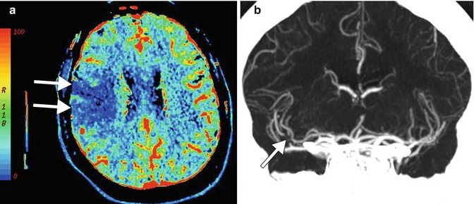
Acute thromboembolic stroke. The patient presented with acute left hemiparesis. CT perfusion imaging (a) demonstrates regional diminished cerebral blood flow within the right MCA territory (arrows). Coronal CTA MIP image (b) demonstrates a focal filling defect within the right MCA superior division (arrow)
Stay updated, free articles. Join our Telegram channel

Full access? Get Clinical Tree


