Acquired Myopathies

76 Acquired Myopathies


Myopathies are disorders that adversely affect muscle function (Greek “myo” = muscle and “pathy” = suffering). Insights from molecular biology are changing the traditional classification of muscle disorders and opening new areas of investigation.


Clinical Vignette


The patient is a 33-year-old woman who presents with a 6-month history of difficulty climbing stairs. Recently she noticed she could not climb the 10 steps from her basement. She has also developed problems picking up jars from shelves of tall cabinets as well as inability to keep her hands over her head to brush her hair. Her ability to swallow has been fine and she has not experienced any shortness of breath. Her examination was significant for bilateral weakness of her hip flexors (iliopsoas), deltoids, biceps, and triceps. Muscle stretch reflexes were diminished but present. Sensory examination was normal.


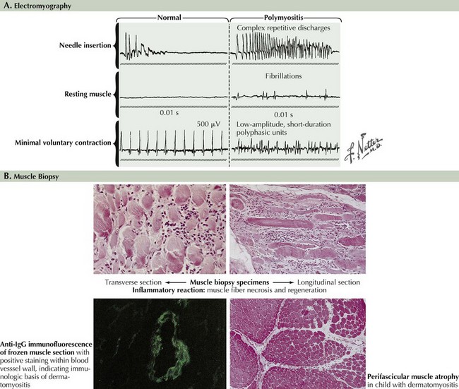
Serum creatine kinase (CK) was increased to 1200 IU/L (six times normal). Electromyography (EMG) demonstrated normal nerve conduction studies. However, needle examination was abnormal, with large numbers of short-duration, low-amplitude, polyphasic motor unit potentials associated with scattered fibrillation potentials and complex repetitive discharges.


Comment: This patient’s presentation is typical for a myopathic process with the clinical picture of evolving proximal weakness, elevated serum CK, and abnormal EMG.


The common nongenetically determined myopathies are classified into those having a primary inflammatory process, an underlying endocrinopathy, a toxic pathophysiology, or an underlying associated systemic disorder. Much less commonly, a few infectious agents, such as trichinosis, may lead to a primary myopathy. Myopathies typically present with symmetric symptoms and signs of muscle weakness affecting the proximal limbs and paraspinal musculature (Fig. 76-1). Asymmetric, distal, generalized, or regional patterns of weakness also occur in certain distinct myopathies such as inclusion body myositis (IBM). Less commonly, ventilatory muscles or cardiac muscles are primarily affected. Myopathies occasionally present with periodic weakness, exercise-induced muscle pain, or stiffness.


Muscle weakness is a common defining feature of a variety of peripheral motor unit disorders. Myopathies are included in the same differential diagnosis as neuromuscular transmission disorders, motor neuron disease, as well as rare demyelinating polyneuropathies. Muscle stretch reflexes are generally normal, and sensation is usually unaffected in primary myopathies. The presence of certain distinguishing clinical features may help in the diagnosis of a myopathy. These include the pattern of weakness (e.g., presence of ptosis, ophthalmoparesis, ventilatory muscle weakness, scapular winging, and head drop) or other clinical features (e.g., contractures, skeletal dysmorphisms, calf hypertrophy, myotonia, cardiac involvement, or subtle to marked dermatologic changes). Another very important diagnostic determinant is an assessment of the clinical temporal profile (e.g., the rate of progression), any history of a relapsing (periodic) weakness, diurnal variation, and symptoms that occur only with exertion. Other important factors include genetic predisposition, medication and toxin exposure, and other organ system involvement.


Diagnostic Approach


Patients who present with symptoms of myalgia and muscle weakness with a normal muscle strength examination and with normal or mildly elevated serum creatine kinase (CK) levels are common in clinical practice. Such patients are diagnostically and therapeutically challenging. Definable myopathic disorders are uncommon in patients who present with muscle pain, fatigue, or exercise intolerance in the absence of objective clinical, laboratory, or electrophysiologic abnormalities.


Laboratory Evaluation


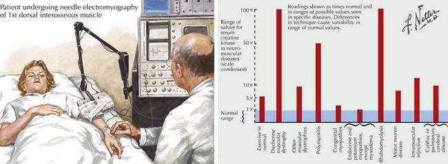
The serum CK is characteristically increased in many myopathies; this may vary from a 2- to 50-fold increase, although in most myopathies CKs are usually in the 500–5000 IU/mL range (Fig. 76-2). When this enzyme is abnormally elevated, its serum levels do not closely parallel disease severity or activity. Serum aldolase levels are also frequently elevated in myopathies; its increase generally parallels the increase in CK, although many clinical neuromuscular specialists do not routinely order an aldolase level. However on occasion it may be elevated with a normal CK as illustrated in the Cushing syndrome vignette reported later in this chapter.


An increased CK level is a nonspecific finding vis-à-vis the diagnosis of myopathies. Other motor unit disorders (such as motor neuron disease, amyotrophic lateral sclerosis, or spinal muscular atrophy) and systemic processes (particularly myxedema) are commonly associated with increased CK of two to five times normal levels. Conversely, the serum CK can be normal in certain patients with DM and IBM.


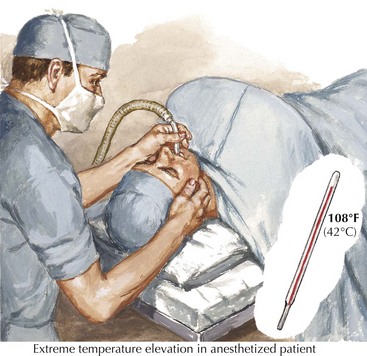
Patients with persistently increased CK levels sometimes associated with muscle pain but without clinically demonstrable weakness, family history, or exposure to potentially myotoxic substances are classified as having hyperCKemia. Despite thorough clinical and laboratory examination, it is often difficult to assign a specific pathophysiologic mechanism to this finding. HyperCKemia is often an elusive clinical challenge. However, it is important to emphasize that although no diagnosis per se is defined, the finding of hyperCKemia deserves serious consideration. Such individuals are at an increased risk of developing malignant hyperthermia (MH) if they require surgery under general anesthesia. Certain induction agents, particularly the halogenated ones, namely halothane, are particularly prone to inducing this life-threatening complication in patients with hyperCKemia. Therefore, we suggest that our hyperCKemia patients wear a MedAlert bracelet to always call the attention of anesthesiologists to this finding and thus potentially prevent an episode of MH (Fig. 76-3).


Serum aspartate and alanine aminotransferases (AST and ALT) are frequently elevated in many myopathies as these enzymes are released by diseased muscle. Rarely some patients with clinically unsuspected myopathies undergo unnecessary evaluation for liver disease when AST and ALT elevations are found and the CK has not been checked. Other liver function studies (e.g., gamma glutamyl transpeptidase and prothrombin time) are normal, providing another clue to the probability of a primary skeletal muscle rather than a hepatic disorder.


Routine biochemistry and hematologic laboratory tests are usually normal in patients with myopathy. Serum potassium levels should be checked to exclude Addison disease with hyperkalemia. Various muscle disorders characterized by episodic periodic paralysis may sometimes have either hypokalemia or hyperkalemia if they are tested during the overt period of paralysis. However, these patients most often have normal potassium values if tested between episodes of weakness. Serum markers of inflammation such as the erythrocyte sedimentation rate (ESR) and C-reactive protein (CRP) may be elevated in some acute myopathies. Thyroid function evaluation (serum TSH levels) must be considered in all patients presenting with an acute or chronic myopathy. Both hypothyroidism and rarely hyperthyroidism may present with primary muscle involvement. Appropriate endocrine evaluation is necessary in myopathic patients when a more obvious diagnosis is not apparent. These also include pituitary adrenal disorders such as Cushing syndrome or Addison disease, and very rarely hyperparathyroidism. In certain ethnic groups, for example, Chinese and Hispanics, thyrotoxicosis may be associated with hypokalemia and a proximal myopathy resembling periodic paralysis.


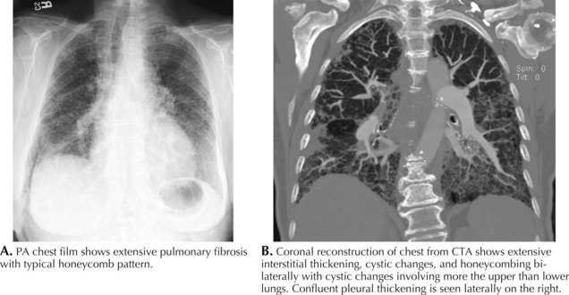
The serum myositis-specific and myositis-associated antibodies are other testing parameters that are useful in the evaluation of some patients with a myopathy. However, as these are present in fewer than half of all patients with polymyositis and dermatomyositis, routine serologic testing for these antibodies is of limited use. The presence of anti-Jo-1 (antibody to histidyl t-RNA synthetase) antibodies suggests potential end organ comorbidity, for example, interstitial lung disease (Fig. 76-4). Signal recognition particle antibodies are most often associated with necrotizing myopathies and may suggest a poor treatment response.


Sometimes polymyositis is associated with an underlying connective tissue disorder. In those patients, serologic markers for the underlying disease may be positive. These include antinuclear antibody (ANA), and/or rheumatoid factor. On occasion, the presence of these antibodies can aid in the diagnosis of the occasional patient for whom the history is not definitive. This is especially so when there is diagnostic confusion between the possibility of an acquired inflammatory myopathy and a genetically determined dystrophy. When polymyositis and, less commonly, dermatomyositis is associated with other collagen-vascular diseases, the combination is referred to as an overlap syndrome. Systemic lupus erythematosus (SLE), systemic sclerosis, rheumatoid arthritis, and Sjögren syndrome may have weakness as a component of their myriad symptoms and signs. In these cases, muscular weakness exceeds what arthritis alone can account for. They are characterized by elevated titers of anti–U1/U2-ribonucleoprotein antibodies, PM-Scl antibodies or SSA antibodies in scleroderma, Sjögren syndrome, SLE, or mixed connective tissue disease. Dermatomyositis is rarely associated with other collagen-vascular diseases, with the exception of scleroderma.


Paraneoplastic antibody evaluation may occasionally be helpful in the differential diagnosis of proximal weakness. This is particularly so in patients with Lambert–Eaton myasthenic syndrome (LEMS) who often present with symptoms emulating a myopathy. These individuals have elevated levels of voltage-gated calcium channel antibodies. This finding, in addition to the classic EMG nerve conduction studies typically seen in LEMS, is very specific for this diagnosis. In addition, anti-Hu antibodies may be positive in patients with myopathy associated with small cell lung cancer.


Immunofixation to look for the presence of serum monoclonal protein is necessary in certain instances if either amyloid myopathy or sporadic late-onset nemaline myopathy (SLONM) is in the differential diagnosis. Approximately 20% of patients with IBM also have a MGUS. Appropriate endocrine evaluation is necessary in myopathic patients when a more obvious diagnosis is not apparent.


Vitamin D levels are also important. Rarely hypovitaminosis D may present with a myopathy. Similarly, patients with primary or secondary osteomalacia may present with proximal weakness. Elevated serum calcium and alkaline phosphatase values may point toward these underrecognized disorders.


Electromyography


EMG evaluation of patients with suspected myopathies is important (Fig. 76-5, and see Fig. 76-2). Results of routine nerve conduction studies are normal in myopathies, with the exception of diminished compound muscle action potential amplitudes in more severe disorders. The primary EMG abnormalities in the myopathies are classically found at the time of the needle examination. Classic findings of a myopathy include the presence of abnormally low amplitude, short duration, and polyphasic motor unit potentials (MUPs). It is typical for these patients to have both an early recruitment and increased numbers of MUPs early on in the muscle activation for a given effort. Destruction of myofibrils or muscle membrane results in abnormal insertional activity, particularly fibrillation potentials and complex repetitive discharges. Inflammatory myopathies, several dystrophies, and various myotonic muscle disorders may be distinguished by the presence of myotonic potentials on needle EMG.


Concomitantly, EMG helps to exclude disorders that affect other anatomic sites within the peripheral motor unit, particularly those with symmetric proximal weakness mimicking a myopathy. These include motor neuron disorders, such as amyotrophic lateral sclerosis, spinal muscular atrophy type 3, chronic inflammatory demyelinating polyneuropathies, neuromuscular transmission disorders (particularly Lambert–Eaton myasthenic syndrome), and myasthenia gravis. Results of EMG are often normal in the various endocrine, mitochondrial, and congenital myopathies.


Muscle Biopsy


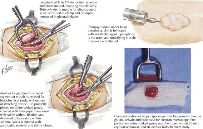
Muscle biopsy is the definitive diagnostic tool for many myopathies (Fig. 76-6). The selection of the biopsy site is important; muscles that are unaffected, that are severely affected (are at end stage), or have been recently subjected to EMG evaluation should be avoided. Muscles commonly biopsied include the vastus lateralis, deltoid, and biceps brachii. The gastrocnemius muscle is often avoided due to the possibility of incidentally discovered neurogenic atrophy. The upper lumbosacral muscles, thoracic paraspinal muscles, such as the multifidus, and much less commonly the cervical paraspinal muscles provide an alternative site for biopsy. On reflection one recognizes that these muscles are indeed the most proximal ones and thus more prone to show early changes of an active myopathic process.


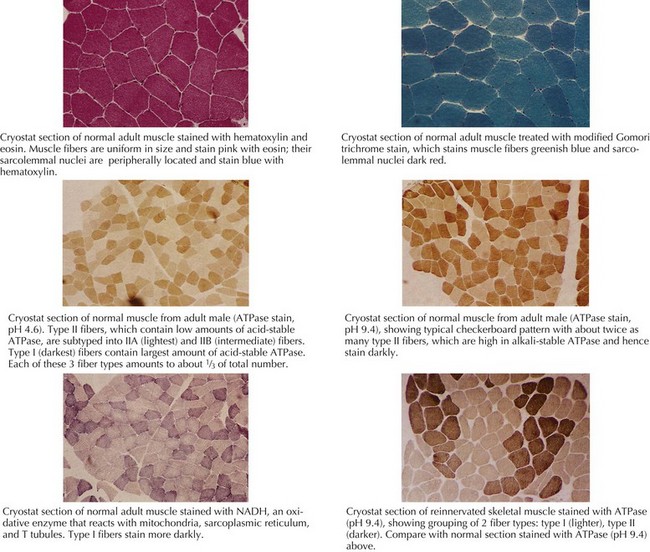
The muscle biopsy specimen per se is divided into separate aliquots for formalin fixation, paraffin embedding, and immediate freezing. The formalin-fixed piece is stained with hematoxylin and eosin (H and E) because this permits a rapid means for initial evaluation. This is especially useful for identifying inflammatory myopathies where such a diagnosis offers the potential for successful therapeutic intervention. Frozen specimens are best for other stains, including nicotinamide adenosine dinucleotide dehydrogenase (NADH), modified Gomori trichrome, adenosine triphosphatase, and lipid and glycogen stains (Fig. 76-7 and see Fig. 76-5).

Muscle biopsy specimens are also subjected to biochemical analysis, mutational analysis, and electron microscopy, when these techniques are indicated. Inherited myopathies, such as the various muscular dystrophies, are evaluated by immunohistochemical stains, immunoblotting; testing is available for calpain, caveolin, dysferlin, the dystroglycans, dystrophin, laminin-2 (formerly merosin), and the sarcoglycans.


Jun 4, 2016 | Posted by in NEUROLOGY | Comments Off on Acquired Myopathies

Full access? Get Clinical Tree

Get Clinical Tree app for offline access