Acquired Polyneuropathies

72 Acquired Polyneuropathies


Diagnostic Approach


Clinical Vignette


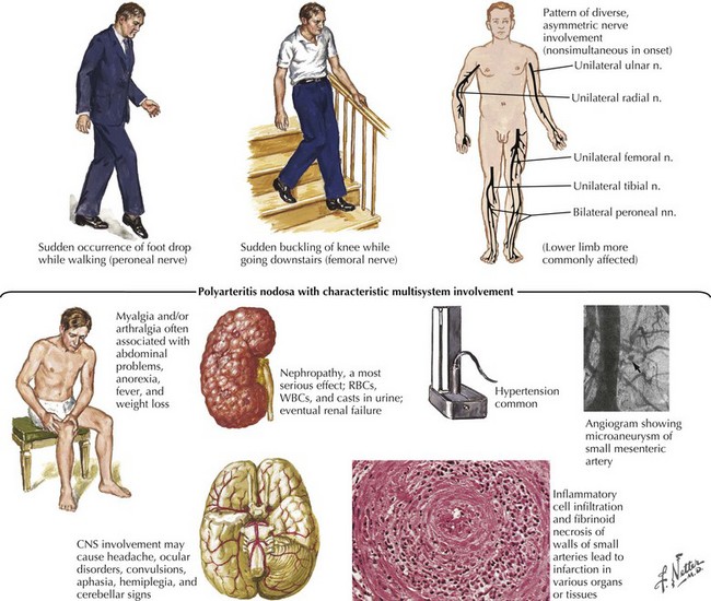
A 51-year-old woman complained of acute onset of right hand weakness and pain for 2 weeks and acute onset of painful left foot drop for 3 days. She describes the pain as being “burning and aching” and associated with some prickling and “pins and needles” sensation. Over the past month she has also experienced low-grade fevers, fatigue, malaise, and an unexplained 15-pound weight loss. Her past medical history is only remarkable for adult-onset asthma, for which she uses an inhaler as needed. She is on no other medications. The family history is unremarkable. She does not smoke or drink alcohol. Review of systems is otherwise negative, including for any symptoms of autonomic nervous system dysfunction.


Her examination was noteworthy for marked weakness and sensory loss in the right ulnar nerve and left peroneal nerve distributions; there was also sensory loss over the lateral aspects of the left foot. Nerve conduction studies (NCS) and needle electromyography (EMG) confirmed moderate to severe right ulnar, left peroneal, and left sural mononeuropathies. NCS/EMG demonstrated relatively recent-onset, prominent axonal damage without any findings of prominent demyelination.


Laboratory testing was remarkable for a very elevated erythrocyte sedimentation rate of 82 mm/hour (nonspecific indicator of systemic inflammation). Hypereosinophilia was present on the complete blood count (CBC). A left sural nerve biopsy was performed confirming the clinical suspicion of vasculitic neuropathy. She was diagnosed with Churg–Strauss syndrome with vasculitic neuropathy and was started on high-dose corticosteroids. This led to gradual resolution of her symptoms; 18 months later, there were only minimal residual deficits in the involved peripheral nerves.


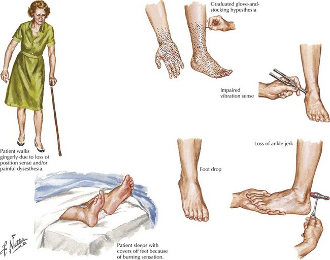
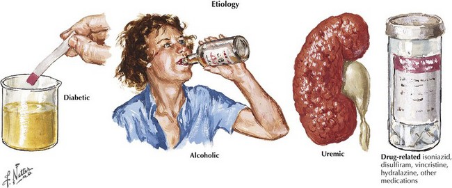
The evaluation for the etiology of a patient’s polyneuropathy can be very challenging for many reasons, including the fact that there are more than 100 potential etiologies (Fig. 72-1). Ultimately the polyneuropathy is determined to be acquired (i.e., caused by some other disease or exposure) in one third of cases (Box 72-1), inherited in another one third of cases (see Chapter 71), and—in spite of appropriate testing—idiopathic in the remaining one third of cases. In order to focus on a smaller list of potential etiologies so that the evaluation can be simplified, we believe that it is best for the clinician to first characterize the polyneuropathy and the patient. We will present one method for characterizing neuropathy that is easy to remember, based on four simple clinical questions about the neuropathy and the patient: “What?” “Where?” “When?” and “What setting?”


“What?” refers to what nerve fiber modalities (motor, sensory, autonomic, or a combination) are involved? The identification of sensory nerve involvement, at a minimum, allows the clinician to exclude other neuromuscular diseases not associated with sensory dysfunction, such as disorders of anterior horn cells (e.g., amyotrophic lateral sclerosis), neuromuscular transmission (e.g., myasthenia gravis), or of muscle (myopathy). When sensory symptoms and signs are present, it is useful to characterize neuropathic sensory symptoms into “positive” or “negative” because acquired neuropathies are usually accompanied by positive neuropathic sensory symptoms and inherited neuropathies are usually not. Positive sensory symptoms may be painful (e.g., “burning,” “freezing,” or “throbbing”) or painless (e.g., “tingling” or “swelling”) (Box 72-2). Paresthesias and pain (positive neuropathic sensory symptoms) are common complaints for patients with diabetic, vasculitic, alcoholic, or uremic neuropathy and patients with Guillain–Barré syndrome (GBS) or chronic inflammatory demyelinating polyradiculoneuropathy (CIDP). In the clinical vignette presented above, the patient had prominent positive neuropathic sensory symptoms, strongly suggesting that the etiology of her neuropathy is acquired rather than inherited. Patients with neuropathy often also complain of exaggerated discomfort to painful sensory stimuli (hyperalgesia) and to nonpainful sensory stimuli (allodynia). Patient complaints indicative of negative neuropathic sensory symptoms include loss of sensation and imbalance (i.e., sensory ataxia). Most patients with neuropathy have some degree of motor nerve involvement that at times is overshadowed by the sensory complaints. Our patient had prominent motor nerve fiber involvement.


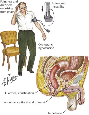
Identification of autonomic nerve involvement can be an important clue because only a small number of neuropathic processes affect both autonomic and somatic nerves (e.g., GBS, paraneoplastic neuropathy, diabetic neuropathy, amyloid neuropathy) (Box 72-3). Autonomic symptoms include lightheadedness, syncope, diarrhea, constipation, postprandial bloating, early satiety, urinary complaints, erectile dysfunction, abnormal or absent sweating, and dry mouth and eyes (Fig. 72-2). Our patient did not have discernible autonomic nervous system involvement.


“Where?” refers to the distribution of nerve damage. An important diagnostic watershed is the determination of whether the process is “length dependent” (e.g., distal) or not. Length-dependent neuropathies manifest first in the feet and are symmetric. Non–length-dependent neuropathies are not necessarily evident initially in the feet and may be asymmetric, focal, or multifocal. The etiology of length-dependent neuropathies is usually inherited, metabolic/toxic, or idiopathic whereas a neuropathy that is not length dependent is often caused by an immune-mediated or infectious process.


Our vignette patient clearly had a neuropathy that was not length-dependent (it was multifocal). Some examples of non–length-dependent neuropathies are polyradiculoneuropathies (e.g., GBS), plexopathies (often inflammatory), sensory ganglionopathy (e.g., paraneoplastic subacute sensory neuronopathy caused by small cell lung cancer), and multifocal mononeuropathies (e.g., mononeuritis multiplex caused by vasculitis). Our patient’s presentation was of painful multifocal mononeuropathies, which is typical of mononeuritis multiplex caused by vasculitis.


“When?” refers to the temporal evolution of the neuropathy. Because of confusion over what is meant by “acute,” “subacute,” and “chronic,” it is often best to describe symptom onset based on whether or not the neuropathic symptoms had a compelling, definite date of onset. A definite date of symptom onset almost always indicates an acute or subacute onset typical of an immune-mediated or infectious etiology. A less-exact date of onset suggests a gradual or insidious onset, indicative of inherited, idiopathic, or toxic/metabolic etiologies. The pace of progression following symptom onset is also an important consideration. Symptom onset and pace of progression often correlate in a predictable manner, owing largely to the underlying mechanism. Mononeuritis multiplex caused by systemic vasculitis, which is at the top of the differential diagnosis for our vignette patient, typically presents with a series of painful mononeuropathies of acute onset, occurring one after the other with the rapid development of significant morbidity. Our patient’s description of acute-onset mononeuropathies is typical of mononeuritis multiplex.


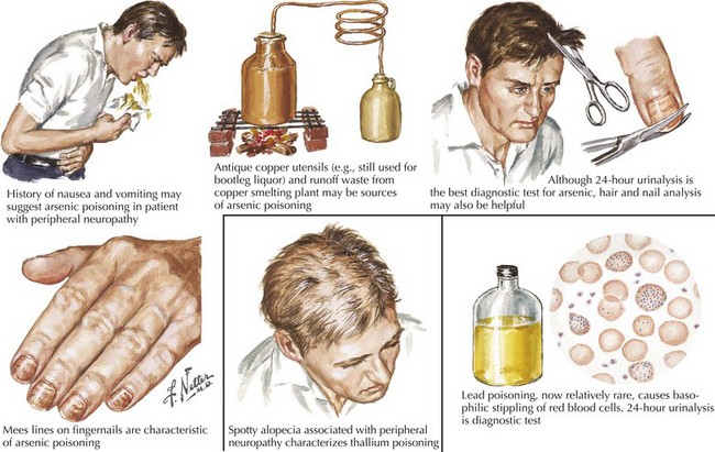
“What setting?” refers to an elaboration of the unique clinical circumstance of the individual patient. This is done by determining what in the patient’s past medical history, medication list, social history, family history, and the review of systems may be relevant (Fig. 72-3). An understanding of the significance of these clinical factors requires knowledge of the risk factors of neuropathy and knowledge of the clinical features of the diseases that may be risk factors for neuropathy. For example, unexplained weight loss raises concern for vasculitis or malignancy (e.g., small cell lung cancer), both of which cause an immune-mediated neuropathy. The neuropathy secondary to malignancy (e.g., paraneoplastic neuropathy) usually presents differently than vasculitic neuropathy, so it is usually not too difficult to differentiate these two etiologies. A clinical setting of known diabetes mellitus or known kidney disease would elevate those comorbidities on the differential diagnosis. Heavy metal poisoning or other intoxication, although rare, needs to be considered in the patient with systemic symptoms (e.g., nausea, vomiting) and other manifestations suspicious for poisoning (Fig. 72-4). Our patient’s presentation is of vasculitic neuropathy rather than paraneoplastic neuropathy.


The fifth step in characterization requires NCS and EMG. NCS and EMG can contribute to (or rarely refute) the clinical characterization in terms of “what” and “where,” as well as provide another view of the temporal evolution (“when”). NCS and EMG can also characterize the neuropathy as being primarily axonal or demyelinating. Neuropathies with axonal injury are far more common than those primarily with demyelination, but the identification of a primarily demyelinating polyneuropathy is very important because acquired demyelinating polyneuropathies (e.g., GBS, chronic inflammatory demyelinating polyradiculoneuropathy, multifocal motor neuropathy) are generally treatable. They are usually immune-mediated and treatable with immunotherapy (e.g., corticosteroids, intravenous immunoglobulin [IVIG], plasmapheresis) (Box 72-4). NCS and EMG can also assess for subclinical involvement and provide baseline parameters in case future testing is necessary to monitor the patient’s course. Our patient’s NCS and EMG confirmed multiple, axonal mononeuropathies. It is important to note that the localization of each mononeuropathy was not at common sites of nerve entrapment, such as the elbow for the ulnar nerve and the fibular head for the peroneal nerve; thus, a compression or entrapment mechanism of injury is not plausible (given the other facts of the case, it would have been very unlikely anyway).


Vasculitis is only one of the more than 100 different causes of neuropathy (Fig. 72-5). It is a far less common cause of acquired neuropathy than diabetic, alcoholic, or uremic neuropathy, but it is very important to diagnose—and to do so quickly—because undiagnosed and untreated systemic vasculitis may be fatal. See discussion on vasculitic neuropathies below.


Clinical Vignette


A 72-year-old man reported a 4-year history of numb feet characterized as a feeling of “cotton between the toes.” Walking on bare feet became uncomfortable. The numbness ascended circumferentially to his ankles. He no longer trusted his balance to put on his pants without support. Difficulty wiggling the toes was the only indication of weakness. He had no symptoms in his hands or face, indications of dysautonomia, or systemic illness. His medications included a diuretic and a multivitamin. He had no toxic exposure or affected family members. The patient appeared well and had bilateral hammertoe deformities. Neurologic findings included an inability to spread his toes and intrinsic foot muscle atrophy. Muscle stretch reflexes were normal in the arms, diminished at the knees, and absent at the ankles. There was a distal maximal graded stocking distribution sensory loss to light touch, pinprick, temperature, vibration, and proprioception to mid calf bilaterally. He wobbled slightly on Romberg testing but did not fall.


EMG demonstrated a length-dependent, primarily axonal, sensorimotor polyneuropathy. An undefined hereditary sensory neuropathy could not be excluded, although his children were examined clinically and electrodiagnostically. Laboratory investigation did not demonstrate an etiologic mechanism. Nerve biopsy was not indicated.


The symmetric pattern of sensory and reflex loss and the subtle distal motor involvement supported the length-dependent nature of his neuropathy. Although his hammertoes were compatible with a hereditary neuropathy, positive sensory symptoms, his age, and the absence of affected family members made Charcot–Marie–Tooth disease (CMT) an unlikely consideration. Distal sensory symptoms could occur with myelopathies; however, the characteristic distribution of clinical findings and absence of urinary sphincter problems was consistent with a length-dependent polyneuropathy (LDPN). Annual follow-up revealed minimal progression of his neuropathy.


Idiopathic Length-Dependent Polyneuropathies


Polyneuropathies are one of the most common neurologic disorders; the length-dependent pattern is the most prevalent. Although there are many recognized causes of polyneuropathy, specific mechanisms have not been identified in 30–40% of patients, and these patients are deemed to have idiopathic neuropathy (Box 72-5).


Box 72-5 Length-Dependent Polyneuropathies


CIDP, chronic inflammatory demyelinating polyradiculoneuropathy; CMT, Charcot–Marie–Tooth neuropathy; GBS, Guillain–Barré syndrome; HNPP, hereditary neuropathy with liability to pressure palsy; LDPN, length-dependent polyneuropathy; MM, mononeuritis multiplex; MMN, multifocal motor neuropathy; PNSS, positive neuropathic sensory symptoms; SN, sensory neuropathy.


Peripheral nerve axons are fine-caliber distal portions of long individual cells, sometimes longer than 1 m. They depend on their cell bodies, within dorsal root ganglia or anterior horns, and their axonal transport mechanisms for nutrition and other factors for maintenance of homeostasis. Length-dependent patterns of dysfunction are thought to relate to impaired cell body metabolism or axonal transport within the nerves’ most vulnerable components. As the neuropathy progresses, the fingers typically become symptomatic when lower extremity symptoms have ascended to approximately the mid-shin to knee level. Very rarely in advanced cases, the chin, nose, and midline trunk are involved.


Typical patients with primary sensory LDPN note tingling, numb, or burning sensations, often pronounced at rest, particularly at night. Occasionally, exercise exacerbates these unpleasant sensations. When weakness is present, foot and toe extensors and foot evertors are more affected than plantar flexors. Gait is compromised because of weakness, or painful dysesthesia. Muscle stretch reflexes are variably affected but commonly ankle jerks are diminished.


Motor-predominant LDPNs have a limited number of etiologies. These are primarily genetically determined (i.e., CMT) or, less commonly, immunologically acquired, multifocal motor neuropathies. More common motor disorders related to diseases affecting the motor neuron (anterior horn cell), neuromuscular junction, or skeletal muscle base usually have asymmetric or generalized patterns of involvement.


Pure sensory neuropathies fit into the LDPN pattern such as with diabetes mellitus, renal disease, vitamin deficiencies, various toxins, Sjögren syndrome, and amyloidosis (Fig. 72-6

Only gold members can continue reading. Log In or Register to continue

Stay updated, free articles. Join our Telegram channel

Jun 4, 2016 | Posted by in NEUROLOGY | Comments Off on Acquired Polyneuropathies

Full access? Get Clinical Tree

Get Clinical Tree app for offline access