13
CHAPTER
![]()
Ambulatory EEG
Robbie D. Buechler
Ambulatory electroencephalography (AEEG) is a monitoring technique that allows the recording of continuous EEG (cEEG) activity when patients are at home. This obviates the need for admission to the hospital for video EEG (vEEG) monitoring in many cases. However, there are situations in which ambulatory EEG monitoring is not adequate, such as in evaluations for surgical treatment of epilepsy. For many other conditions in which spells need to be characterized, this type of monitoring may be sufficient. Details on the background, principles, and importance of EEG in general are described in Chapter 11.
HISTORICAL PERSPECTIVE
From the humble beginnings and discovery of EEG by German neurologist Hans Berger in 1924, technology and smaller and more high-powered computers have dramatically advanced this field (1). The evolution of EEG into an ambulatory monitoring system first came about via technology put forth by cardiologist, Dr. Holter. Holter was an American biophysicist who invented the Holter monitor, a portable device for continuously monitoring the electrical activity of the heart for 24 hours or more. He donated the rights to his invention to medicine, and his namesake continues on in describing this long-term ECG monitoring system. Soon, buffer delay memory systems based on event monitoring and push button capture of episodes, similar but less sophisticated than ones used for in-patient vEEG monitoring, were incorporated into ambulatory monitoring.
The initial ambulatory system was an EEG preamplifier added to Holter’s 4-channel, 24-hour ECG monitoring system. The first person to wear an AEEG did so in Montreal in 1973. In 1982, a 16-channel AEEG system was introduced that utilized signal multiplexing (2). The 16 channels allowed improved spatial resolution and localization, but recorded discrete samples rather than cEEG. In 1983, a cassette tape AEEG system was introduced. It used off-head preamplifiers that had continuous 8-channel recording capability, real-time event markers, and gain and filter adjustments (2,3). Unfortunately, most of our knowledge about the sensitivity, specificity, and pitfalls of AEEG comes from older, technically limited studies (4). Some of these are detailed later. Renovating this outdated data with up-to-date studies using new more powerful, complex, and even video-containing systems is desperately needed.
In the past decade, computer technology has enabled portable recording of up to 36 channels with sampling rates of up to 400 Hz. Currently, numerous AEEG systems are available commercially. The addition of video monitoring has once again advanced the field and has helped negate one of the biggest disadvantages of AEEG – the lack of visualization of the episodes (5). There is a limited amount of comparative data on the effectiveness of AEEG in contrast to other techniques. This is especially true of newer systems, which have vEEG capabilities.
TECHNICAL CONSIDERATIONS
Due to the active environment during an AEEG recording, disk electrodes are applied with collodion, the patient’s scalp wrapped in gauze, and the lead wires tacked to reduce the chance of disconnection of the electrodes. Examples of commercially available AEEG equipment are shown in Figures 13.1A and B.
Patients are given detailed instructions on recording daily activities and events in a log, as well as in how to use the push-button activation for notable episodes. Though battery life is a concern, with extra batteries, a patient description sheet, and careful demonstration on how to use the equipment, a 72-hour study can be performed without much issue.
Currently available 16- to 36-channel AEEG systems use the similar amplifiers as used for in-patient vEEG monitoring systems, except that many do not have video input. Some also have video capability, but none has the maximal channel (64–128) capability of sophisticated in-patient wired systems. However, these additional channels are not typically needed, as the standard EEG montages are adequate for most uses. New AEEG machines reduce reviewing time by means of sampling (which risks missing infrequent discharges) and automated spike and event detection programs (2).

FIGURE 13.1A Shoulder harness style-AEEG without video.
![]()
Despite the progress made with AEEG systems, there remain some technical limitations. Because it is obtained in a very active environment, AEEGs can be fraught with artifacts. Sometimes artifacts can look like electrographic seizures. Electroencephalographic seizures usually “tell a story” with a beginning, evolution with neighborhood/local spread, and an ending, including postictal suppression. Artifacts do not tell such a story. Knowledge and experience can often differentiate true versus artifactual paroxysmal events. Movement, electromyographic, and other types of artifact can also make interpretation of data difficult. Ideally, patient log entries, filtering, and montage selection can ameliorate some of these technical limitations. The data storage limitations of older devices have largely been ameliorated with technological updates to computer processors and ambulatory storage. Archiving of data still needs to be well planned to provide adequate storage, just as in in-patient vEEG monitoring.
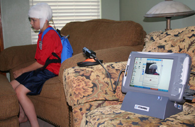
FIGURE 13.1B Backpack-style AEEG with separate video recorder; wide-angle vEEG one piece systems are also available (not shown).
![]()
APPLICATIONS AND INDICATIONS
The Neurophysiology Subcommittee of the ILAE recommends the use of long-term monitoring where the diagnosis of epilepsy or the classification of the seizure syndrome is difficult to establish (5). This guidance also states that “ambulatory outpatient and community-based long-term monitoring may be used as a substitute for inpatient long-term monitoring in cases where the latter is not cost-effective or feasible or when activation procedures aimed at increasing seizure yield are not indicated.” At this time, there are no specific guidelines or practice parameters for AEEG; however, there are three main reasons why AEEG is performed: a) to characterize behavior events, b) to better define seizure type and epilepsy syndrome, and c) to obtain a longer sample to capture interictal epileptiform discharges (IEDs).
Behavioral Events
Perhaps the most common reason to perform AEEG is typically when clinical evaluation and baseline EEG are not adequate for diagnosis of epilepsy and if nonepileptic spells are suspected. Objective data are instrumental in the differential diagnosis of epilepsy from psychogenic nonepileptic seizures (PNES), syncope, parasommias, cardiac arrhythmias, transient ischemic attacks, or other behavioral disturbances.
PNES are the most common type of spell confused with epilepsy. A clinical diagnosis of epilepsy is found to be incorrect in many patients, with many of the incorrectly diagnosed patients having PNES. Many cases of PNES are due to conversion disorder, anxiety, depression, or a combination of these. AEEG may be particularly effective in capturing stress-related PNES as the patient is at home under typical conditions with the usual life stressors. Comparatively, if the monitoring is performed as an in-patient, the typical home stressors are absent, and stress-related spells may be less likely to occur. PNES are discussed in detail elsewhere in this text.
Syncope, particularly convulsive syncope, may be difficult to clinically differentiate from seizures. AEEG has been used in this situation to determine if seizures may be the cause of syncope. Epileptic abnormalities are unusual in patients with syncope, near-syncope, or episodes of dizziness (6). Most syncopal episodes are nonneurological and do not occur with the frequency that allows them to be captured on AEEG.
Nocturnal events have also been studied with AEEG. Sleep is more natural in the home environment than in the hospital, so AEEG is well suited for this determination. Absence of video is somewhat limiting in this situation, but if epileptiform abnormalities are present, the diagnosis is clear. However, the absence of epileptiform abnormalities does not necessarily rule out frontal lobe seizures, and also makes parasomnias a possibility as well.
Characterization of Seizures and Epilepsy
Even if a diagnosis of epilepsy is clear, AEEG can be helpful in further clarifying seizure type (ie, focal versus generalized) or epilepsy syndrome. In some cases, seizures are considered “intractable” to medications, but, in reality, incorrect AEDs have been used – generalized seizures being treated with medications for focal seizures. Similarly, correct syndromic diagnosis can provide additional information about genetics, prognosis, and treatment that allows more effective treatment.
Interictal Epileptiform Discharges
A routine EEG is typically 20 to 30 minutes. This is a relatively short window in which to capture transient discharges like IEDs that may help make a diagnosis of epilepsy. With new AEEG systems, EEG data are stored for the entire recording time rather than just during push button alarms or during seizures. The increased EEG sampling time to 48 to 72 hours increases the probability that IEDs will be captured in patients with epilepsy (7). With AEEG studies, natural sleep is captured as well, which also enhances the ability to capture IEDs. Because of these potential advantages in capturing IEDs, AEEGs have been used not only in the diagnosis of epilepsy but also to see if AEDs can be safely withdrawn. Whether AEEG actually helps in making AED withdrawal safer has not been confirmed.
Once recordings extend beyond 72 hours (sometimes even 48 hours), electrode contact starts to deteriorate and artifacts increase. This makes recognizing IEDs more difficult, though electrographic seizures may still be evident. Moreover, combing through a 20- to 30-minute routine EEG for IEDs is commonly done; however, diligently evaluating each page of a 72-hour recording for IED is much more difficult. Automated spike detectors currently lack the sensitivity of seizure detectors, and so they cannot be relied on to identify IEDs.
Other Indications
There may be other reasons to obtain an AEEG as well. At times, AEEG has been used to determine seizure frequency. This is particularly helpful in situations in which the patient or caregiver is uncertain of the seizure frequency and the provider suspects frequent episodes (8). Alternatively, there may be subtle clinical episodes that are not reliably recognized by the patient and provider. AEEG, especially when combined with video, can help quantify the number of such episodes during the length of the recording. In some situations, AEEG has been used in the evaluation for epilepsy surgery (9). This is not done very often as AEDs cannot be tapered and detailed video analysis is necessary for determination of seizure semiology. In this situation, simply determining that a spell has epileptic correlate is not adequate – the electrographic onset must be determined. If data quality is compromised, as it often the case later in the AEEG recording, the onset may not be clearly identifiable.
UTILITY AND COMPARISON TO OTHER TYPES OF STUDIES
A true comparison of AEEG to in-patient vEEG monitoring is difficult due to lack of well-controlled studies using modern-day AEEG equipment. Older studies are limited because they used four to eight channels of AEEG. Some studies evaluate the diagnostic yield by whether IEDs are detected, while others look at the presence of typical spells. Still other studies compare AEEG to a routine EEG. This leaves anecdotal uncontrolled smaller studies and case reports to fill in some of the enormous gaps left by these outdated studies. A recent review nicely summarizes prior studies (7).
In one study in which pediatric patients were evaluated, AEEG was able to capture and determine the nature of spells in 61% of patients undergoing this test for spell characterization (10). For children being evaluated for frequency of IEDs or to better characterize seizure type, AEEG was successful in every case. Another study that prospectively evaluated patients for spell characterization, diagnosis of epilepsy, IED counting, or to determine frequency of seizures, AEEG provided “useful information” in 72% of patients (11). In an adult study in which data were evaluated slightly differently, “positive data” were obtained in 68% of patients (12). Positive data included capturing spells and IEDs. Video recording has been shown to increase the diagnostic yield of AEEGs by 25% to 45%, especially in patients with frequent paroxysmal events (13). Adding video to AEEG, of course, would not be expected to increase utility in cases in which only frequency of IEDs needs to be determined.
Inpatient vEEG monitoring can be inconclusive in determining the nature of spells if the spell is not captured and if IED are not identified. Patients having such nondiagnostic vEEG monitoring studies were discharged with AEEG in one study (14). The diagnostic yield with the AEEG increased by 33% for these patients.
AEEG has shown utility in determining seizure frequency as well. Often patients and caregivers may be unaware of the frequency of seizures, especially when they occur in sleep or when the patient is alone. In one study, patients self-identified only 62% of ictal events with an additional 38% identified by AEEG (8). In 23% of patients, seizures were detected by the program but not recognized by the patient. That is, the patient had a seizure but was not aware of having a seizure. Lack of awareness and underreporting of seizures have significant clinical consequences. A patient’s ability to drive will depend on their ability to recognize their seizures. Individuals with unexplained injuries may benefit from AEEG since it may help identify seizures as the cause of the injuries.
The yield of AEEG in comparison to baseline EEG is considerably higher. Baseline EEG has relatively low sensitivity in detecting IEDs, and often multiple studies must be done. With AEEG, up to 78% of selected patients had IEDs detected (10). However, in another study of children and adults, IEDs were seen in only 26% of patients, but this still shows an improvement over routine EEG (15). An advantage of AEEG is the diurnal recording. When IEDs occur primarily soon after awakening from sleep, they can be more easily recorded with AEEG than with routine EEG. In one series, almost 5% of 1,000 consecutive patients were noted to have generalized IED soon after awakening from sleep, helping confirm their diagnosis of juvenile myoclonic epilepsy (16).
The duration of AEEG has also been investigated. In one study, 96-hour AEEG assisted with the diagnosis of spells as epileptic or not in 51% of patients (12). Of the events recorded, 58% were recorded within 24 hours and 78% within 48 hours. From these data, 42% and 22% of first events would have been missed if the recording was only for 24 or 48 hours, respectively.
ADVANTAGES AND DISADVANTAGES
Advantages
AEEG has many advantages when compared to inpatient vEEG monitoring. Low cost and convenience are probably the biggest advantages. However, there are other benefits as well.
Ambulatory EEG is a cost-effective alternative to inpatient vEEG monitoring in some situations (11). Costs are estimated at 51% to 65% lower than a 24-hour inpatient admission for vEEG monitoring and up to 90% less when monitoring lasts for more than 3 days (16).
The convenience of AEEG is very attractive. Its outpatient nature circumvents the need for hospitalization. Family and caregivers do not need to take time away from their schedules to be with the patient in the hospital. Keeping the patient out of the hospital also does not reduce mobility (which may have other consequences) and prevents exposure to nosocomial infections. For ambulatory patients who are employed or are in school, they may continue going to work or school while undergoing AEEG.
With AEEG, capturing the effects of the patient’s natural environment is possible. Various aspects of the environment, such as physical activity, stress, and heat often provoke seizures and spells. These natural spell precipitants often cannot be replicated when the patient is hospitalized. In the home environment, sleep is more natural and not as disrupted as in the hospital. Natural sleep may provoke abnormalities that may help make the diagnosis (17). Recording EEG during the entire natural environmental circadian cycle is useful in capturing spells that occur only at certain times of the day. In addition, circadian variations of IEDs are well documented.
Disadvantages
Along with the many advantages of AEEG, there are some disadvantages. One of the most significant disadvantages is lack of technologist’s support in the nonhospital environment. Because the patient is ambulatory, the probability that electrodes will be dislodged is high and increases as the duration of recording increases. This can introduce artifacts that may make interpretation impossible. Even if electrodes are not dislodged, desiccation of the gel or paste that enables electrode contact with skin can compromise recording fidelity. Some laboratories circumvent this by having patients return for maintenance of electrodes after 48 to 72 hours. This also allows downloading acquired data and providing the patient with new batteries for the AEEG unit.
Another important limitation is lack of video recording. Absence of video makes determining spell semiology difficult. This raises the issue of whether the spell captured was indeed the patient’s typical episode. Sometimes artifacts can mimic electrographic seizures. The presence of video may make this distinction easy, and its absence can raise more questions or lead to misinterpretation. Some newer AEEG units, as noted earlier, have video capability. But even these newer units have a stationary video camera that is usually set in one location. It may not be recording the patient at the moment of the spell, thus defeating its purpose. Nonetheless, having a camera at least increases the odds of capturing the semiology.
One of the common methods of inducing seizures in patients hospitalized for vEEG monitoring is tapering their AEDs. This cannot be done with AEEG. Medication taper may result in a flurry of seizures or tonic–clonic seizures necessitating intervention. This cannot be done in an outpatient, at home setting. This further limits the utility of AEEG.
FUTURE DIRECTIONS
The utility of AEEG will continue to improve as technology improves. Better battery life and greater storage capacity will allow units to run longer and capture longer samples of EEG. This may require electrode maintenance during the study. Better computer algorithms to detect seizures and IEDs will reduce the human error in identifying epileptic abnormalities. Improved video capabilities may enable better video integration into AEEG machines. Improvements in electrodes may allow for better skin contact and better quality studies. However, what are needed most are high quality studies comparing the utility of AEEG (with and without video) to routine EEG and inpatient vEEG monitoring. This will help practitioners decide which study is most appropriate for their patients.


