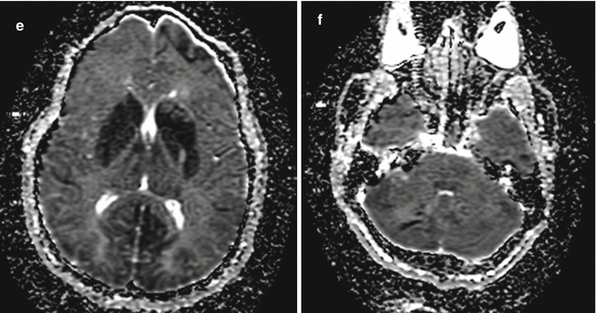
Fig. 6.1
Acute Ecstasy toxicity. Axial FLAIR (a, b), DWI (c, d), and ADC map (e, f) show multiple supratentorial and infratentorial acute infarcts. Courtesy of Alisa Gean

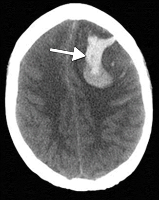
Fig. 6.2
Ecstasy-induced hemorrhage. The patient presented with acute severe headache immediately after “two hits” of Ecstasy. Axial CT image demonstrates a large left frontal lobe intraparenchymal hematoma (arrow)

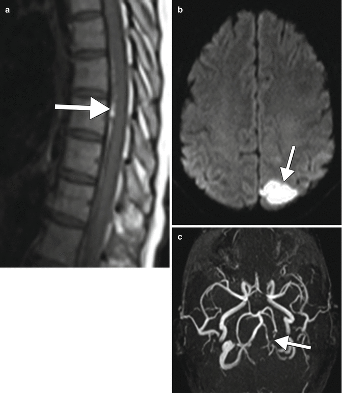
Fig. 6.3
Methamphetamine-induced hemorrhage, infarct, and vasospasm. Sagittal T1 MRI of the spine (a) reveals intradural spinal hemorrhage (arrow). No intracranial subarachnoid hemorrhage was evident. Axial diffusion-weighted image (c) reveals a left occipital lobe infarct (arrow). MRA MIP image (b) reveals multiple areas of vasoconstriction, most pronounced in the left PCA (arrow). The patient was treated with supportive care, and angiography performed 10 weeks later (not shown) confirmed resolution of vasoconstriction
6.4 Differential Diagnosis
Reversible cerebral vasoconstriction syndrome (RCVS): RCVS encompasses a diverse group of conditions characterized by the acute onset of severe headaches associated with reversible segmental constriction of multiple cerebral arteries. Importantly, the underlying pathophysiology is reversible vasoconstriction rather than vasculitis. Of note, it is not possible to distinguish between RCVS and CNS vasculitis based on imaging. However, several clinical features are helpful in making the diagnosis. This is essential, as treatment and prognosis of RCVS and CNS vasculitis are quite different. The onset of RCVS is acute with thunderclap headache. However, the severe pain is short lived, lasting from a few minutes to a few hours. Attacks can be single, but patients have a mean of four attacks during a period of approximately 1–4 weeks. A self-limiting, benign outcome is the norm, and resolution is generally within 3 months. CSF analysis is usual normal or near normal. The neuroimaging hallmark of RCVS is vasoconstriction evident as segmental narrowing and dilatation (string of beads) of one or more arteries (Fig. 6.4). The appearance is similar to that of other forms of vasculitis, except the abnormalities resolve within a few months. Bilateral and diffuse anterior and posterior circulation changes are seen in the majority of patients. Although concomitant changes need not be present, subarachnoid hemorrhage, parenchymal edema, parenchymal hemorrhage, and infarction may occur. Of note, RCVS may be accompanied by posterior reversible encephalopathy syndrome (PRES), as these are not mutually exclusive. RCVS patients are treated with nimodipine or verapamil until resolution of angiographic findings.

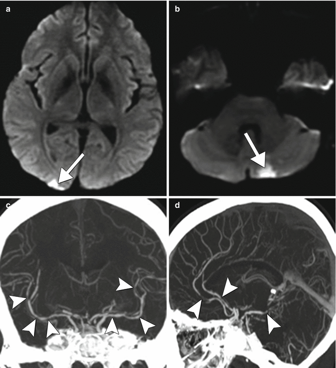
Fig. 6.4
Reversible cerebral vasoconstriction syndrome (RCVS). The patient presented with acute onset of thunderclap headache in the setting of Prozac and Cannabis use. Axial DWI (a, b) images demonstrate acute areas of infarction (arrows). Coronal (c) and sagittal (d) CTA reconstructions demonstrate multifocal areas of arterial narrowing and irregularity involving the anterior, middle, and posterior cerebral arteries (arrows). CSF analysis was normal and symptoms resolved within 3 months
Stay updated, free articles. Join our Telegram channel

Full access? Get Clinical Tree


