Amyotrophic Lateral Sclerosis

67 Amyotrophic Lateral Sclerosis


Her examination revealed normal cognitive function. Cranial nerve examination revealed mild dysarthria, tongue fasciculations, the presence of a jaw jerk, and weakness of neck flexion. All limb muscles were weak, left more than right, more pronounced distally. Intrinsic hand muscles were atrophic. Fasciculations were noted throughout her limbs. Muscle stretch reflexes were brisk despite her weakness and atrophy. Plantar responses were extensor. Sensory examination revealed no abnormalities.


In 1874, Jean-Martin Charcot described a disorder that he named amyotrophic lateral sclerosis (ALS). In France, it is referred to as Charcot disease, whereas motor neuron disease (MND) is the preferred name for the disorder in the United Kingdom. In the United States, ALS is better known as Lou Gehrig’s disease.


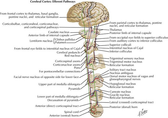
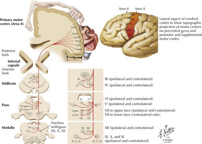
Charcot’s description was of a disorder characterized by loss of voluntary motor function, resulting from degeneration of anterior horn cells, corticospinal tracts, and motor cranial nerve nuclei and cortical motor neurons (Figs. 67-1 and 67-2). ALS is a sporadic disorder (sALS) in the majority of cases. ALS is inherited in 5–10% of cases, i.e., familial ALS (fALS), usually in an autosomal dominant fashion. In general, fALS patients have phenotypes that closely resemble sALS, although fALS may have an earlier onset. In absence of family history, the disorders are clinically indistinguishable.


The incidence of ALS approximates 1.8 in 100,000. The incidence of ALS in men is twice that in women, although this ratio becomes closer to 1 : 1 in a postmenopausal population. The median age at onset is 55 years of age; this disease may afflict patients in their late teens or in their 90s. The average life expectancy is between 2 and 3 years; in less than 10% of patients, ventilator-independent survival of less than 1 year or greater than 10 years is seen. Half of affected individuals die within 3 years and only a quarter survive 5 years without dependency on invasive mechanical ventilation. Young males and patients with restricted upper motor neuron (UMN) or lower motor neuron (LMN) presentations tend to have a slower course. Primary bulbar (disordered speech and swallowing) presentations tend to disproportionately affect older women and appear to have a more rapid course.


In the United States, it is estimated that at any given time 25,000 patients are diagnosed with ALS. The prevalence of ALS appears to be increasing, perhaps because of an aging population. Other than historical observations identifying an increased incidence on Guam and the Kii peninsula of Japan, there does not appear to be any particular geographic location or ethnic group that has a significantly higher risk of contracting ALS.


Etiology, Genetics, and Pathogenesis


The cause of sporadic ALS is unknown. As with other neurodegenerative diseases, it is hypothesized that ALS may result from the dual insult of genetic susceptibility and environmental injury. Attempts to identify predisposing mutations and potential toxic or infectious agents have been unsuccessful to date.


It has been long recognized that a small percentage of patients (fALS) have an autosomal dominant pattern of Mendelian inheritance. A major breakthrough in our understanding of familial ALS took place in 1991 with the identification of cytosolic copper-zinc superoxide dismutase (SOD1) gene mutations on chromosome 21q22.11 in affected individuals in some families. This represents the most frequently identified form of fALS. SOD1 is a free radical scavenger. Recognition of the SOD1 mutation led to the hypothesis that SOD1-fALS was mediated by free radical toxicity. However, SOD1 knock-out mouse with no SOD1 protein do not develop motor neuron disease. In contrast, heterozygote mice become symptomatic and die from a paralyzing disorder. It is thought that SOD1 mutations may injure neurons through conformational changes in the SOD1 protein.


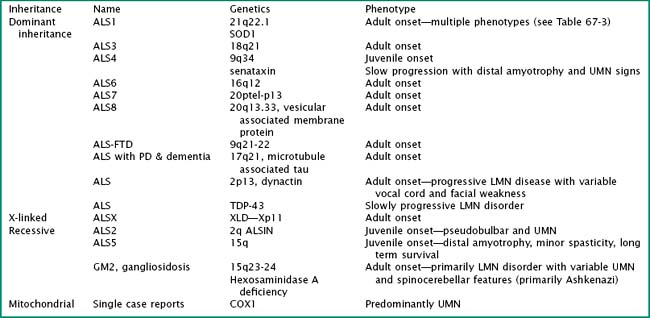
Particularly intriguing has been the recognition of the phenotypic heterogeneity in SOD1 fALS (Table 67-1). About 114 pathologic mutations have been identified within the five exons of the SOD1 gene; each of these mutations may produce a distinct phenotype. The most common mutation found in North America is an alanine for valine (A4V) substitution at codon 4; this typically produces a lower motor neuron dominant phenotype (LMN-D) with a life expectancy approximating 1 year. Table 67-1 summarizes the phenotypic heterogeneity that results from different SOD1 mutations. SOD1 mutations are not fully penetrant. It is estimated that individuals carrying the mutation have an 80% chance of developing disease by age 85 years. SOD1 mutations constitute 20–25% of all individuals with fALS. Other fALS genotypes are listed in Table 67-2. Some of these mutations produce a predominantly lower motor neuron (LMN) or upper motor neuron (UMN) disorder and more closely resemble the phenotypes of spinal muscular atrophy or hereditary spastic paraparesis, respectively.


Table 67-1 Phenotypic Variation in SOD1 fALS


Phenotype SOD 1 Mutation
Lower motor neuron predominant A4V, L84V, D101N
Upper motor neuron predominant D90A
Slow progression (>10-year survival) G37R, G41D, G93C, L144S, L144F
Fast progression (<2-year survival) A4T, N86S, L106V, V148G
Late onset G85R, H46R
Early onset G37R, L38V
Female predominant G41D
Bulbar onset V148I
Low penetrance D90A, I113T
Posterior column involvement E100G


Mutations that may produce both a frontotemporal lobar degeneration and motor neuron disease occur on chromosomes 9p13.2-21.3, 9q21-q22, and 17q21. A recently identified fALS mutation occurs in the TAR DNA-binding protein, 43 (TDP-43) gene. Non-amyloid, structurally modified TDP-43 has been recognized as a major constituent of the ubiquitinated inclusions found in cortical neurons of patients with both sporadic (s) and familial (f) forms of frontotemporal lobar degeneration (FTD).


There are many proposed mechanisms for motor neuron death in sALS, including excitotoxicity secondary to glutamate, free radical–mediated oxidative cytotoxicity, mitochondrial dysfunction, protein aggregation, cytoskeletal abnormalities, aberrant activation of cyclo-oxygenase, impaired axonal transport, activation of inflammatory cascades, and apoptosis. Why the motor neurons and corticospinal/bulbar tracts are vulnerable in a selective manner remains unknown. Why the disease begins focally and progresses in a regional fashion is also unknown. One putative hypothesis is that misfolded, toxic protein aggregates may proselytize normal protein in adjacent neurons, analogous to mechanisms proposed for prion diseases.


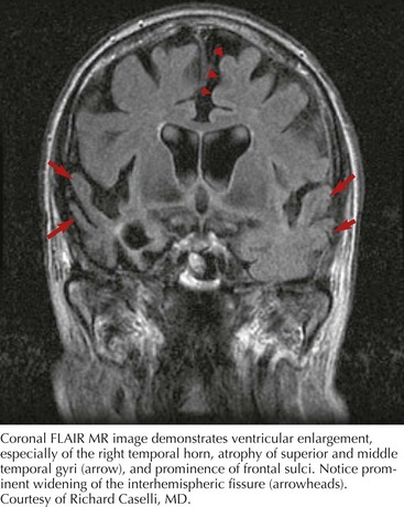
Whatever the mechanism, ALS is pathologically characterized by loss of myelinated fibers in the corticospinal and corticobulbar pathways (see Fig. 67-2) and loss of motor neurons within the anterior horns of the spinal cord and many motor cranial nerve nuclei. Even in individuals with predominantly UMN or LMN involvement clinically, pathologic involvement of both systems is seen. Patients with associated FTD have preferential lobar atrophy and neuronal loss from these portions of the brain (Fig. 67-3). As a result of anterior horn cell loss, ventral roots become atrophic in comparison to sparing of their dorsal root counterparts (Fig. 67-4). Anterior horn cell loss occurs within virtually all levels of the spinal cord with selective sparing of the third, fourth, and sixth cranial nerves, and Onuf’s nucleus within the anterior horn of sacral segments 2–4. There is also cell preservation within the intermediolateral cell columns.


The majority of sALS patients will be found to have ubiquitinated inclusions and Bunina bodies within the central nervous system. The latter are dense granular intracytoplasmic inclusions within motor neurons considered specific for ALS. Additionally in ALS with FTD, spongiform changes of the first and second layers of the frontal cortex have been described.


Clinical Presentations


The diagnosis of ALS remains a clinical endeavor. EMG and nerve conduction studies and measurements of ventilatory capacity are routinely obtained in ALS suspects. These tests are done to provide diagnostic support for diffuse LMN and ventilatory muscle involvement respectively. Other testing is done with the primary intent of identifying or excluding differential diagnostic considerations. SOD1 mutational analysis provides diagnostic proof in patients with suggestive family histories. Although a small percentage (2%) of seemingly sALS patients will be found to have SOD1 mutations, mutational analysis is not routinely recommended in this population.


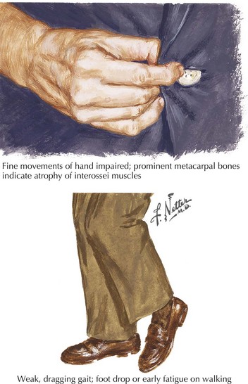
The presenting features of ALS are quite variable. Typically, the patient seeks medical care when his or her weakness begins to affect activities of daily living (Fig. 67-5). It is not uncommon for ALS to be misdiagnosed initially and the time between symptom onset and diagnosis is usually months. Unfortunately, there is a tendency to misdiagnose ALS as a potentially treatable nerve, nerve root, or spinal cord compressive syndrome or orthopedic condition. A significant percentage of ALS patients may undergo unnecessary surgeries. It should be emphasized that progressive weakness and atrophy in the absence of pain and sensory symptoms rarely represents a surgically treatable condition.


The exclusive motor involvement and the chronologic course serve to distinguish ALS from other neurologic disease. Simultaneous involvement of both UMNs and LMNs and progression both within and outside of the originally involved regions is necessary for a definite clinical diagnosis. LMN involvement may be documented by clinical, electrodiagnostic, or pathologic (muscle biopsy) means. UMN involvement is currently defined by clinical criteria alone. Classic ALS is usually an easy diagnosis for an experienced neurologist. Diagnosis may be delayed in patients with limited clinical evidence of the LMN or UMN involvement, slow disease progression, and confounding neurologic signs from unrelated problems such as sensory loss from mononeuropathies, radiculopathies, and polyneuropathies.


The signature of anterior horn cell loss is painless weakness and atrophy, hypoactive or absent deep tendon reflexes, and fasciculations. Atrophy is best appreciated when it is focal, in contrast to normal muscle bulk elsewhere. It may be difficult to distinguish atrophy from LMN disease resulting from the atrophy of disuse, particularly in the elderly. Suppressed deep tendon reflexes may also be difficult interpret as they may represent a normal variant. In ALS, muscle weakness due to LMN dysfunction occurs in a segmental (myotomal) distribution and spreads in a regional fashion. As an example, a patient with ALS and hand weakness may have all hand muscles innervated by C8–T1 roots affected. Weakness occurring in a nerve distribution should lead to consideration of a different disorder, for example, multifocal motor neuropathy.


Fasciculations seen in many muscles in multiple limbs are ominous and are strongly suggestive of a motor neuron disease. Fasciculations that occur infrequently, or repetitively in one area, are more likely to have a benign origin, particularly in the absence of weakness or atrophy. The absence of fasciculations does not eliminate ALS. They may not be readily visible because of prominent subcutaneous tissue. Physicians often initially recognize fasciculations, although the patient in retrospect may recall that they were present for some time. Muscle cramping is a common, albeit nonspecific, manifestation of motor neuron disease. The initiation of cramps during manual muscle testing is common in ALS.


What constitutes clinical signs of corticospinal or corticobulbar tract pathology may be more ambiguous. Spasticity represents a definite UMN sign; Babinski signs if unequivocal are confirmatory of UMN disease. Unfortunately, Babinski signs may not be elucidated in many ALS patients as a result of LMN toe extensor weakness. The Hoffman sign is generally indicative of UMN involvement of the arms, particularly if asymmetric. Hyperactive deep tendon reflexes, particularly with sustained clonus, indicate UMN involvement. The term relative UMN sign has been used to describe the presence of a deep tendon reflex in a weak and atrophic muscle. Reflex spread also implicates UMN disease; finger flexion (C8) occurring in response to brachioradialis tendon percussion (C6), and activation of the contralateral thigh adductors when the insertions of the ipsilateral thigh adductors are percussed (crossed adduction) are two notable examples of this phenomenon. Motor impairment in UMN disease typically results in slowness and incoordination. UMN weakness occurs in a specific pattern: the elbow, wrist, and finger extensors are weaker than their flexor counterparts in the upper extremity whereas conversely, hip and knee flexors and foot dorsiflexors and evertors are weaker in the lower limb.


Tongue atrophy, fasciculation, and weakness are perhaps the most frequently occurring and recognized manifestations of LMN involvement of cranial nerves. It is important to observe for fasciculations when the tongue is relaxed on floor of the mouth (Fig. 67-6). Tremulousness of the tongue with attempted protrusion may be readily misinterpreted as representing fasciculations. Weakness of both facial and jaw muscles may occur in ALS but they are usually subtle. Weakness of neck extension and neck flexion is common in ALS, and head drop may be a rare presenting feature (Fig. 67-7). Neck drop is commonly associated with posterior neck discomfort and is typically relieved when the neck is supported. Notable for their absence are ptosis and ophthalmoparesis, and symptoms related to sight, hearing, taste, smell, and facial sensation.

Only gold members can continue reading. Log In or Register to continue

Stay updated, free articles. Join our Telegram channel

Jun 4, 2016 | Posted by in NEUROLOGY | Comments Off on Amyotrophic Lateral Sclerosis

Full access? Get Clinical Tree
Get Clinical Tree app for offline access