(1)
Department of Anesthesiology and Intensive Care Medicine, Neckar-Odenwald Clinic, Dr. Konrad-Adenauer-Strasse 37, 74722 Buchen, Germany
Abstract
Airway maintenance in patients with obstructive sleep apnea (OSA) can be challenging; therefore, profound preoperative evaluation and preparation are mandatory. Decisions for anesthesiologic airway management focus on awake fiberoptic intubation vs. induction of general anesthesia, and on the preservation of or the return to spontaneous ventilation. The strategies differ based on preoperative knowledge of intubation difficulties and airway abnormalities. The recovery phase after the end of the surgery calls for the same alertness as the initial management of airway difficulties. Close cooperation of surgeon and anesthesiologist in the pre, intra, and postoperative period will help reduce airway-related morbidity and mortality.
Core Features
Airway maintenance in patients with obstructive sleep apnea (OSA) can be challenging; therefore, profound preoperative evaluation and preparation are mandatory.
Decisions for anesthesiologic airway management focus on awake fiberoptic intubation vs. induction of general anesthesia, and on the preservation of or the return to spontaneous ventilation.
The strategies differ based on preoperative knowledge of intubation difficulties and airway abnormalities.
The recovery phase after the end of the surgery calls for the same alertness as the initial management of airway difficulties.
Close cooperation of surgeon and anesthesiologist in the pre, intra, and postoperative period will help reduce airway-related morbidity and mortality.
13.1 Implications in Patients with OSA
Obstructive sleep apnea (OSA) is, by definition, a problem of the upper airway. Its presence indicates an increased likelihood of difficult intubation and airway maintenance under anesthesia. Three different groups of patients undergoing general anesthesia can be defined, each requiring different strategies for managing the airway: patients who have been diagnosed for OSA, patients with symptoms suggesting sleep apnea, and patients who lack signs of the syndrome or in whom such features are missed preoperatively. Surgical procedures can be OSA-related or for any other diagnosis with varying invasiveness. The common goal in all cases will be to avoid inadequate ventilation and oxygenation resulting in hypoxemia or hypercarbia and any associated hemodynamic changes (e.g., tachycardia, arrhythmia, and hypertension) leading to increased morbidity and mortality. Death, brain injury, and cardiopulmonary arrest, are amongst the most severe adverse events associated with difficulties in airway management, with airway trauma and damage to teeth as additional risks [106].
Widespread guidelines for management of the difficult airway have been introduced by the American Society of Anesthesiologists (ASA) in 1992 and have been published in revised form in 2003 [20]. A difficult airway is defined as the clinical situation in which a conventionally trained anesthesiologist experiences difficulty with face mask ventilation, difficulty with tracheal intubation, or both. The purpose of the ASA guidelines is to reduce the likelihood of adverse outcomes. the main goal is to ensure adequate oxygenation whenever difficulties occur. As D.B. Scott wrote more than 20 years ago: patients do not die from “failure to intubate,” but from failure to stop trying or from undiagnosed esophageal intubation [654].
The first step in managing the difficult airway is to identify any patient at risk in the preoperative period. Evidence on prediction of difficult airways is inconclusive, but any patient with a diagnosed OSA or presenting clinical signs such as obesity, limited mouth opening or a large tongue should be treated as a patient with a difficult airway until proven otherwise. The description of a difficult airway in previous anesthesia records or in an anesthesia pass issued by an anesthesiologist offers clinically suggestive evidence that the difficulty may reoccur. Whenever possible, an airway history should be obtained prior to the initiation of anesthetic care in all patients. Focused medical history, physical examination, and review of medical records may improve the detection of a difficult airway in patients with OSA, therefore enabling the anesthesiologist to prepare the patient and the anesthesia team for the occurrence of airway difficulties. In some patients, additional evaluation may be indicated to further judge the likelihood of the anticipated airway difficulty.
It is widely accepted that preparatory effects will help in minimizing the risk in patients presenting with a difficult airway. In case of a known or suspected difficult airway, the patient should be informed of the special risks and procedures for management of the difficult airway. An additional experienced anesthesiologist should be available, specialized equipment for difficult airway management should be prepared, and a strategy as well as a backup strategy based on an institutional algorithm for establishing a secure airway should be defined (Fig. 13.1). Preoxygenation should be performed for 3 or more minutes and supplemental oxygen should be administered whenever possible during the process of establishing a secure airway and also after extubation.
Fig. 13.1
Algorithm “airway management”
There are several basic management choices when faced with a suspected or known difficult airway: awake intubation vs. induction of general anesthesia followed by intubation attempts, the preservation vs. the ablation of spontaneous ventilation, noninvasive vs. invasive techniques for the initial approach to intubation.
13.2 Strategies for Intubation in Patients with a Known or Suspected Difficult Airway
One important strategy in patients with a known or suspected difficult airway may be the avoidance of the necessity for invasive airway management. Whenever feasible, local anesthesia infiltration or regional blockades should be preferred in these patients.
In patients with OSA, awake intubation may be attempted with a fibreoptic bronchoscope after application of local anesthetics. Once the glottis has been successfully identified, the tracheal tube can be advanced and general anesthesia can be inducted. If the procedure fails due to lack of patient cooperation, difficulties in identifying the glottic aperture caused by anatomical aberrations or massive secretion, the attempt may be canceled, the feasibility of local or regional anesthesia may be considered, or the decision for invasive airway access may be made. The latter might be considered as first choice in some patients with the options of surgical or percutaneous tracheostomy and surgical or needle cricothyrotomy with the option of jet ventilation.
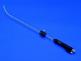
When general anesthesia is induced in patients with a known or suspected difficult airway, tracheal intubation will still be successful in a number of patients without problems, especially when performed by an experienced anesthesiologist. If the initial intubation attempt fails, an additional anesthesiologist should be called if not already present, and the option of awakening the patient and returning to spontaneous ventilation should be considered based on the urgency of the surgical procedure. The possibility of face mask ventilation is the key issue for all further proceedings: when face mask ventilation and oxygenation of the patient are possible, repeated attempts at tracheal intubation can be attempted, with modified techniques where necessary. Alternative approaches may be the use of a fibreoptic bronchoscope, possibly in combination with a supraglottic airway device, the use of rigid fiberscopes such as the Bonfils [58] (Fig. 13.2), of special larnygoscope blades such as the McCoy-blade with the option of additional leverage of the epiglottis (Fig. 13.3), or the use of an intubation stylet. The Bonfils allows overcoming a major problem in many OSA patients, passage of the big tongue and visualization of the vocal chords even when the epiglottis cannot be lifted sufficiently with the laryngoscope. This device has become an integral part of managing airway difficulties in our department ever since it was introduced. In many patients, correct positioning of the head will further aid in achieving the goal of tracheal intubation. Pressure on the cricoid cartilage by an assistant, ideally backward, upward, and toward the right (“BURP” – backward, upward, rightward pressure) may also facilitate identification of the glottis. Any “blind” intubation attempts without visualization of the glottis should be avoided for they may lead to trauma and swelling, interfering with further management of the airway.