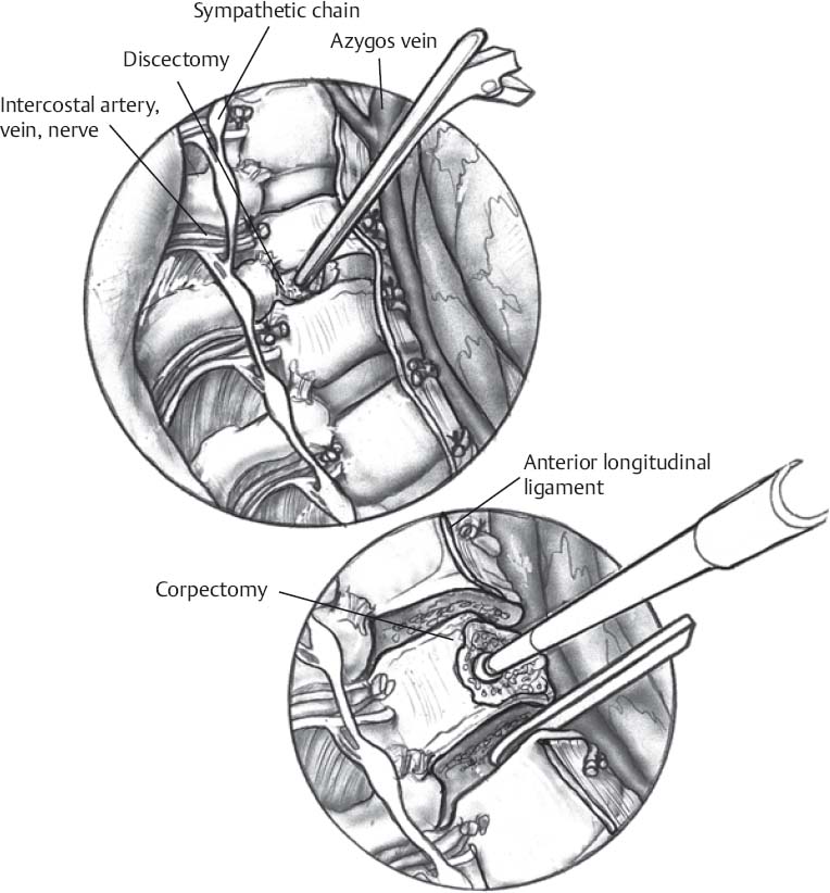
♦ Preoperative
Imaging
- Magnetic resonance imaging (MRI) to assess spinal cord compression and extent of pathology (soft tissue and pleural extension). Important to determine to what extent ventral compression is based on the disc spaces versus behind the bodies. Computed tomography (CT) scan to identify or confirm calcified disc or other ossified pathology.
- Plain x-rays to evaluate alignment, count ribs, and identify natural fiducials which may facilitate correlation of intraoperative imaging findings with MRI images
Preoperative Care
- Approach as described in Chapter 105, Transthoracic Thoracotomy.
- Preoperative plan should also have target anatomic loci which can correlate intraoperative images with preoperative MRI findings
- Review of imaging important to determine extent of decompression that will be required. A clear preoperative plan is useful (e.g., T7–T8 disc space versus caudal half of T7 and rostral one third of T8 versus T6–T7 to T7–T8 and behind the entire body of T7).
Equipment
- Self retaining thoracic (rib) retraction system
- Extended length Bovie may be useful
- Long handled Kerrisons, pituitaries, and curettes
- Long Frazier suction tips may be useful
- Drill with long attachments and bits may be useful
< div class='tao-gold-member'>
Only gold members can continue reading. Log In or Register to continue
Stay updated, free articles. Join our Telegram channel

Full access? Get Clinical Tree


