Approach to the Patient with Neurogenic Orthostatic Hypotension, Sexual and Urinary Dysfunction, and Other Autonomic Disorders


Approach to the Patient with Neurogenic Orthostatic Hypotension, Sexual and Urinary Dysfunction, and Other Autonomic Disorders


Emilio Oribe

Bhuwan P. Garg


The autonomic nervous system (ANS) maintains internal homeostasis and regulates protective responses by continuously monitoring and responding to internal and external stimuli. This is achieved through autonomic reflex pathways and extensive vasomotor, visceromotor, and sensory innervation. The baroreflex, an example of an autonomic reflex, regulates blood pressure (BP), heart rate (HR), and extracellular fluid volume (Fig. 31.1).

ANS dysfunction may be focal or generalized depending on the site of a lesion in an autonomic reflex pathway. Central forms of ANS dysfunction are because of lesions involving neurons of the CNS, brainstem, spinal cord, and preganglionic neurons, whereas with peripheral forms, dysfunction is because of lesions involving peripheral ganglia and postganglionic neurons, or to lesions involving afferent autonomic reflex limbs (Fig. 31.1). Selected autonomic symptoms and findings are summarized in Table 31.1. Numerous diseases result in autonomic dysfunction (Table 31.2).


I. EVALUATION OF THE PATIENT WITH ANS SYMPTOMS


A. History.

All patients presenting with autonomic dysfunction should undergo a com prehensive medical and neurologic history and physical examination. Important elements of the autonomic history include the following:


1. Chief autonomic complaints,

with severity of symptoms, their distribution and frequency, progression, the presence of aggravating and alleviating factors, and a measure of the degree of disability are determined.


2. Review of ANS systems

including cardiovascular, sexual, urinary, gastrointestinal, vasomotor, thermoregulatory and sudomotor, secretomotor, pupillomotor, and sleep functions.


3. Medication review

(antihypertensive, psychotropic, antiandrogenic, laxative medications, and alcohol and recreational drugs can produce ANS dysfunction).


4. Psychosocial evaluation

to determine the impact of ANS dysfunction on quality of life.


5. Family history

(inherited autonomic disorders).


B. Physical examination.

The physical and neurologic examination indicates the site and extent of the lesion responsible for ANS dysfunction and defines associated illness. A comprehensive examination includes supine and upright BP and HR, and examination of the skin and mucosa to assess sweating patterns and to determine if trophic lesions are present. Avascular system examination is important. In patients with genitourinary and anorectal dysfunction complaints (see the following list), the physical examination also includes the following:

1. Abdominal examination to determine if aortic dilatation is present (1% of patients with erectile dysfunction [ED] have abdominal aortic aneurysm) or masses are present.

2. Stretching and palpating the penis for an indication of the integrity of erectile tissue and if Peyronie’s plaques (lumps within the penis) are present.

3. Testicular volume and consistency.

4. Cremasteric reflex (testicle retraction on stroking the thigh), anal wink reflex (anal sphincter contraction on stroking perianal skin), and bulbocavernosus reflex (anal sphincter contraction on squeezing the glans penis or clitoris).

5. Rectal examination to determine if prostatic hypertrophy, fecal impaction, and prolapse are present.


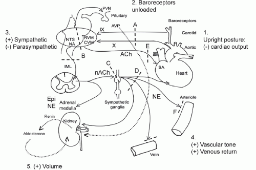
FIGURE 31.1 The baroreflex. 1. Standing produces “pooling” of 600-1,000 mL of blood to the lower body (mainly limb and splanchnic capacitance circulation), reducing venous return and cardiac output (by approximately 30%). 2. This is sensed by specialized stretch receptors (arterial and cardiopulmonary baroreceptors) that in turn activate (unload) baroreflexes. Inputs from carotid and aortic baroreceptors travel with the glossopharyngeal (IX) and vagus (X) nerves to converge on cardiovascular centers in the brainstem and medulla (mainly nucleus of the tractus solitarius) and their projections. 3. The physiologic baroreceptor reflex response to the volume shifts produced by upright posture is a compensatory increase in sympathetic tone with a decrease in parasympathetic outflow. 4. Sympathetic nerve terminals release NE that produces increased vasoconstriction of skeletal and mesenteric muscle vessels, HR, and cardiac contractility. Additional increases in venous return occur through a “pumping” effect of contracting limb and abdominal muscles engaged by the effort of standing (not shown). 5. A longer term response promoting extracellular fluid volume expansion includes baroreflexmediated release of renin, angiotensin, and aldosterone, leading to increased renal Na++ absorption, and release of vasopressin, with an increase in free-water absorption. A: Afferent baroreflex limb dysfunction is present when lesions involve baroreceptors and IX and X cranial nerves (neck surgery, radiotherapy, trauma, neuropathies, and autonomic disorders such as baroreflex failure, Holmes-Adie syndrome, and HSAN III). B: Central lesions involve the ventrolateral medulla (MSA), descending sympathetic pathways (medullary lesions, spinal cord lesions above T5), and IML (MSA, Lewy’s body disorders). C: Sympathetic ganglia lesions involve sympathetic ganglia (autoimmune ganglionopathies associated with nAChR antibodies or paraneoplastic, Lewy’s body disorders). D, E: Postganglionic sympathetic and efferent parasympathetic lesions are present with small fiber neuropathies (diabetes, amyloidosis, Sjögren’s syndrome, HSAN III). F: Efferent sympathetic neuroeffector junction dysfunction occurs with dopamine βhydroxylase deficiency and α-1 adrenoceptor blocking drugs. nAChR, nicotinic acetylcholine receptor antibodies; AVP, arginine vasopressin; CVM, caudal ventrolateral medulla; Epi, epinephrine; HSAN III, hereditary sensory autonomic neuropathy type III; IML, intermediolateral cell columns of the spinal cord; NA, nucleus ambiguus; NE: morepimephrime NTS, nucleus tractus solitarius; RVM, rostral ventrolateral medulla; SA, sinus node.


TABLE 31.1 Clinical Features of ANS Dysfunction


Common Symptoms


Examination


Cardiovascular autonomic


Orthostatic intolerance: syncope, near-syncope, lightheadedness, confusion and impaired cognition, weakness, slurring, visual disturbances, tremors, neck and shoulder (coat hanger) aches


OH, hypotension, orthostatic tachycardia


Exercise intolerance, silent myocardial infarction, intraoperative cardiovascular liability, increased mortality


Tachycardia, loss of HR respiratory sinus arrhythmia. Intraoperative hypothermia, reduced hypoxic-ventilatory drive


Vasomotor


Skin discoloration


Flushing, pallor, distal cyanosis, trophic changes. Changes in skin blood flow absent


Sudomotor and thermoregulatory


Hypohidrosis, hyperhidrosis, heat/cold intolerance


Abnormal temperature regulation, hypothermia, hyperthermia


Secretomotor


Dry mouth, excessive salivation, gustatory sweating, tearing, dry eyes


Dry mouth, dry eyes


Gastrointestinal


Difficulty swallowing, constipation, early satiety, bloating, nausea, vomiting, abdominal pain/cramping, diarrhea, fecal incontinence, weight loss


Abnormal bowel sounds, abdominal distention, reduced anal tone. Gastroparesis, esophageal enteropathy


Bladder


Incontinence, urgency, weak stream, dribbling, hesitancy, retention, recurrent infection


Distended bladder, increased postvoid residual


Sexual


Loss of libido, decreased genital engorgement, ED, ejaculatory dysfunction, dyspareunia


Decreased penile or clitoral and labial engorgement, decreased genital lubrication


Pupillomotor


Glare, blurred vision, poor night vision


Impaired pupillary responses


C. Autonomic testing.

ANS testing is considered an extension of the physical examination. The data from ANS testing is of most value when the selection of tests is guided by the clinical findings. The goal of testing is to confirm the presence of autonomic dysfunction, determine the extent of autonomic involvement, and to localize the site of a lesion in the ANS reflex arc, and distinguish primary from secondary autonomic disorders. Most ANS tests assess the integrity of a reflex arc by recording stimulus-evoked effector organ responses. The more popular tests rely on measuring those effector organ responses that are easily recorded (i.e., changes in HR, BP, pupillary size, etc.). Common bedside tests are shown in Table 31.3. More sophisticated tests include beat-to-beat arterial pressure recordings during different challenges, and the measurement of sweat output (the quantitative sudomotor axon reflex test [QSART], measuring efferent sudomotor function). Usually a battery of several tests is required to reach a diagnosis of autonomic dysfunction (abnormal tests do not always imply disease).


1. Screening tests.

Selection of testing is directed by the clinical presentation.


  • a. Complete blood count and differential.


  • b. Urinalysis and renal function studies.


  • c. Hemoglobin A1C, fasting and postprandial glucose, and glucose tolerance test.


  • d. TSH levels.


  • e. HIV testing.


  • f. Immunoelectrophoresis (serum and urine).


  • g. Sweat gland nerve fiber density and epidermal nerve fiber density (skin biopsy).


  • h. Aminolevulinic acid, porphobilinogen, and porphyrins (24 hour urine collection), erythrocyte porphobilinogen deaminase activity.


  • i. Genetic testing (inherited neuropathies).


  • j. Amyloid staining in fat aspirate, rectal or gingival biopsy.


  • k. [Norepinephrine] plasma (supine and standing)


TABLE 31.2 Selected Autonomic Disorders


Autonomic Disorders


Central autonomic disorders


MSA


PAF (peripheral involvement predominant), Parkinson’s disease, DLB


Disorders of different causes (cerebrovascular, epileptic, tumoral, demyelinating, traumatic, infectious, and degenerative) and autonomic presentations involving primarily: frontal lobes, limbic system, hypothalamus, brainstem, cerebellum, and spinal cord.


Autonomic neuropathies


a. Acute and subacute autonomic neuropathies: subacute autoimmune autonomic neuropathy (panautonomic neuropathy and pandysautonomia), subacute paraneoplastic autonomic neuropathy, Landry-Guillain-Barré’s syndrome, botulism, porphyrias, drug induced and toxic autonomic neuropathies.


b. Chronic peripheral autonomic neuropathies: distal small fiber neuropathies.


Sympathetic and parasympathetic neuropathies: DAN, amyloidosis, autoimmune autonomic neuropathy (paraneoplastic and idiopathic), sensory neuronopathy with autonomic failure, hereditary neuropathies.


Catecholamine disorders


Baroreflex failure, tumors that secrete catecholamines (pheochromocytoma, neuroblastoma, chemodectoma, and familial paraganglioma syndrome), disorders affecting neurotransmitter metabolism (tetrahydrobiopterin deficiency, aminoacid decarboxylase deficiency, dopamine β hydroxylase deficiency and Menkes’ disease, monoamine oxidase deficiency states, and dopamine metabolism disorders).


Orthostatic intolerance disorders


POTS


Mitral valve prolapse dysautonomia


Idiopathic hypovolemia


Paroxysmal syncope


Neurally mediated hypotension and bradycardia (vasovagal), situational syncope


Miscellaneous


Hyperhidrosis (generalized, focal), anhidrosis (CNS, peripheral nerve, and dermatologic)


Horner syndrome, Holmes-Adie’s syndrome, Ross’ syndrome, and crocodile tears


Hirschsprung’s disease


Brugada’s syndrome


Abbreviation: DAN, diasetic autonomic meuropathy; DLB, dementia with Lewy bodies; PAF, pure autonomic Failure; POTS, postural tachycardia syndrome.


D. Antibody testing.

Should always be guided by the clinical presentation.

1. Antinuclear antibodies, rheumatoid factor, Anti-Ro/SS-A, and Anti-La/SS-B.

2. Other antibodies: Neuronal nicotinic acetylcholine receptor, P/Q-type calcium channel, and acetylcholine receptor.

a. Paraneoplastic antibodies: Anti-Hu (ANNA-1); Purkinje-cell cytoplasmic antibodies type 2 (PCA-2); and collapsin response-mediator protein 5 (CRMP-5), voltage gated calcium channels (VGCC).


TABLE 31.3 Bedside Autonomic Tests


Response


Test


Methods


Normal


Abnormal


Site of lesion


Active standing (supine rest for >3 min, or until BP, HR stable followed by unaided standing)


BP and HR supine and standing after 3 min


↑HR<30 beats/min


= HR*


Sympathetic efferents


↓ SBP <30mm Hg


↓ SBP


HRmax/HRmin during first 30 s of active standing (30:15 ratio)


> 1.02


1.00


Vagal efferents


Deep breathing (6 breaths/min)


Maximum—minimum HR


> 10 beats/min


<8 beats/ min


Vagal efferents


Maximum R-R in expiration/minimum (E/I ratio)


> 1.10


< 1.05


Valsalva’s maneuver (blow into a mouthpiece maintaining 40 mm Hg for 15 s)


Longest R-R after maneuver/shortest R-R


> 1.15


< 1.10


No rebound bradycardia in phase IV: vagal efferents


No tachycardia in phase II: S efferents


Mental stress (independent of baroreflex afferents)


HR and BP 2 min into the stress of mental arithmetic (i.e., serial 7 subtractions for 2.5 min)


↑ HR>12


Sympathetic efferents


NE release in response to upright posture


Supine and upright plasma NE


=


Sympathetic efferents


Autonomic tests are used to explore the integrity of whole or of portions of autonomic reflex arcs. Abbreviation: R-R, electrocardiographic R-R intervals.


Exaggerated increases in HR suggest hypovolemia, deconditioning, POTS. Bedside tests require a sphygmomanometer and ECG or EMG instruments (typical settings: low-frequency filter =1-5 Hz and high-frequency filters =500 Hz). Plasma NE determinations are available through commercial laboratories. Testing should be performed in a quiet, comfortable environment.


E. Electrophysiologic studies.

Nerve conduction studies and EMG help define large fiber peripheral neuropathies. Sphincter and pelvic floor EMG is a specialized technique (useful when performed by experienced examiners) in detecting denervation potentials in selected muscles in lesions of the anterior horn cells in the spinal cord.


F. Imaging.

MRI of the brain and spine is essential in CNS and spinal cord lesions. Pelvic imaging may be indicated in those in whom structural lesions are suspected.


Clinical Presentation of Autonomic Dysfunction


NEUROGENIC ORTHOSTATIC (POSTURAL) HYPOTENSION

Orthostatic hypotension (OH) is a frequent and disabling manifestation of autonomic disorders. It may be the initial sign (i.e., “tip of the iceberg”) heralding the onset of primary and secondary autonomic disorders.


A. Diagnosis.

BP measured with a sphygmomanometer and pulse rate recorded while supine for a few minutes of quiet rest (i.e., once BP values have stabilized), and after standing up for 3 minutes is sufficient to determine if OH is present and may help with its differential diagnosis (Table 31.4). OH is defined as a sustained decrease in systolic pressure of at least 20 mm Hg (30 mm Hg in patients with supine hypertension) and of diastolic pressure of at least 10 mm Hg within 3 minutes of standing or head up tilt to at least 60 degrees on a tilt table.


B. Pathophysiology.

The autonomic responses to gravitational volume shifts are com plex, and rely on intact baroreflexes (Fig. 31.1). In neurogenic OH, a patient’s ability to normally increase vascular tone in upright postures is impaired as a result of a failure to appropriately release norepinephrine (NE), the sympathetic postganglionic neurotransmitter innervating blood vessels. This may be because of an impaired afferent baroreflex pathway or to impaired efferent sympathetic outflow at central or peripheral sites. In OH, the lesions are below the medullary circulation centers.

Only gold members can continue reading. Log In or Register to continue

Stay updated, free articles. Join our Telegram channel

Aug 18, 2016 | Posted by in NEUROLOGY | Comments Off on Approach to the Patient with Neurogenic Orthostatic Hypotension, Sexual and Urinary Dysfunction, and Other Autonomic Disorders

Full access? Get Clinical Tree

Get Clinical Tree app for offline access