Fig. 8.1
Betel nut-induced oral cavity squamous cell carcinoma. Axial (a) and coronal (b) contrast-enhanced CT images show an enhancing mass in the right oral tongue (arrows)

Fig. 8.2
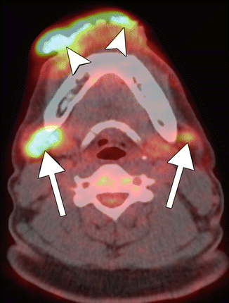
Inflammatory lymph nodes associated with betel nut-induced squamous cell carcinoma. Axial PET-CT fusion image shows an exophytic hypermetabolic lower lip squamous cell carcinoma (arrowheads) and bilateral hypermetabolic level 1 lymph nodes (arrows) that did not contain malignant cells
8.4 Differential Diagnosis
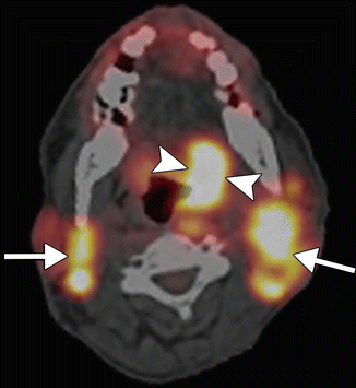
Oral region squamous cell carcinomas can be predisposed by other factors besides betel nuts, including tobacco, alcohol, and HPV (refer to Chaps. 1 and 2). The imaging features of squamous cell associated with betel nut use generally appear similar to other squamous cell carcinomas on imaging. However, HPV positive squamous cell carcinomas tend to be well defined with cystic metastatic lymph nodes. In addition, betel nut-induced squamous cell carcinomas are particularly frequently associated with enlarged, hypermetabolic inflammatory lymph nodes. The false-positive inflammatory lymph nodes have been shown to have maximum standard uptake values (mSUV) on 18FDG-PET that are indistinguishable from metastatic lymph nodes (Fig. 8.3). Lymph nodes may also exhibit hypermetabolism on PET in the weeks following radiation therapy, but the SUV tends not to be as elevated.


Fig. 8.3
Metastatic lymph nodes. Axial PET-CT fused image shows a hypermetabolic left tongue base squamous cell carcinoma (arrowheads) and bilateral hypermetabolic metastatic lymphadenopathy (arrows)


