5 Blood supply of the brain
Arterial Supply of the Forebrain
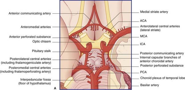
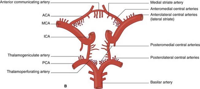
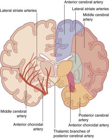
The blood supply to the forebrain is derived from the two internal carotid arteries and from the basilar artery (Figure 5.1).
Anterior cerebral artery (Figure 5.2)
The anterior cerebral artery passes above the optic chiasm to gain the medial surface of the cerebral hemisphere. It forms an arch around the genu of the corpus callosum, making it easy to identify in a carotid angiogram (see later). Close to the anterior communicating artery, it gives off the medial striate artery, also known as the recurrent artery of Heubner (pron.‘Hoibner’), which contributes to the blood supply of the internal capsule. Cortical branches of the anterior cerebral artery supply the medial surface of the hemisphere as far back as the parietooccipital sulcus (Table 5.1). The branches overlap on to the orbital and lateral surfaces of the hemisphere.
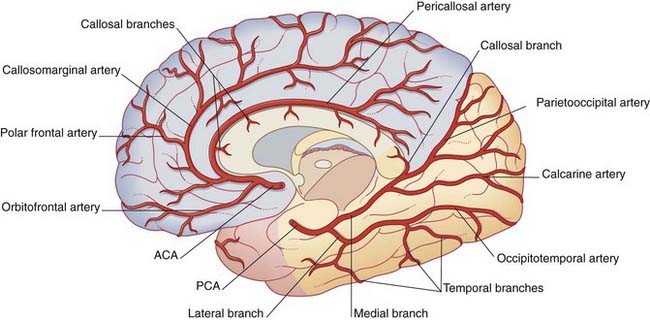
Table 5.1 Named corticala branches of the anterior cerebral artery
| Branch | Territory |
|---|---|
| Orbitofrontal | Orbital surface of frontal lobe |
| Polar frontal | Frontal pole |
| Callosomarginal | Cingulate and superior frontal gyri; paracentral lobule |
| Pericallosal | Corpus callosum |
a The term cortical is conventional. Terminal is better, because these arteries also supply the underlying white matter.
Middle cerebral artery (Figure 5.3)
The middle cerebral artery is the main continuation of the internal carotid, receiving 60–80% of the carotid blood flow. It immediately gives off important central branches, then passes along the depth of the lateral fissure to reach the surface of the insula. There it usually breaks into upper and lower divisions. The upper division supplies the frontal lobe, the lower division supplies the parietal and temporal lobes and the midregion of the optic radiation. Named branches and their territories are listed in Table 5.2. Overall, the middle cerebral supplies two-thirds of the lateral surface of the brain.
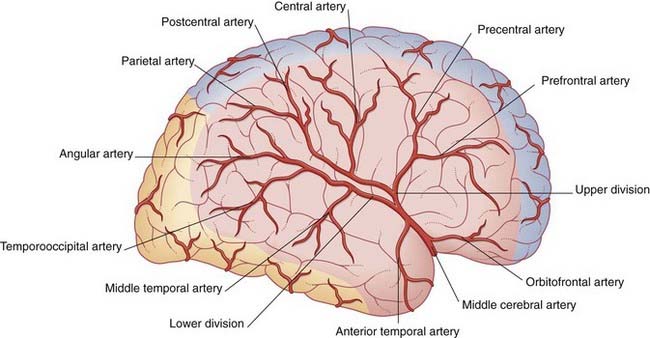
Table 5.2 Cortical branches of the middle cerebral artery
| Origin | Branch(es) | Territory |
|---|---|---|
| Stem | Frontobasal | Orbital surface of frontal lobe |
| Anterior temporal | Anterior temporal cortex | |
| Upper division | Prefrontal | Prefrontal cortex |
| Precentral | Premotor areas | |
| Central | Pre- and postcentral gyri | |
| Postcentral | Postcentral and anterior parietal cortex | |
| Parietal | Posterior parietal cortex | |
| Lower division | Middle temporal | Midtemporal cortex |
| Temporooccipital | Temporal and occipital cortex | |
| Angular | Angular and neighboring gyri |
The central branches of the middle cerebral include the lateral striate arteries (Figure 5.4). These arteries supply the corpus striatum, internal capsule, and thalamus. Occlusion of one of the lateral striate arteries is the chief cause of classic stroke, where damage to the pyramidal tract in the posterior limb of the internal capsule causes hemiplegia, a term denoting paralysis of the contralateral arm, leg, and lower part of face.
Note: Additional information on the blood supply of the internal capsule is provided in Chapter 35.
Posterior cerebral artery (Figures 5.2 and 5.5)
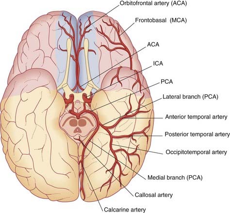
Close to its origin, each posterior cerebral artery gives branches to the midbrain and a posterior choroidal artery to the choroid plexus of the lateral ventricle. Additional, central branches are sent into the posterior perforated substance (Figure 5.1). The main artery winds around the midbrain in company with the optic tract. It supplies the splenium of the corpus callosum and the cortex of the occipital and temporal lobes. Named cortical branches and their territories are given in Table 5.3.
Table 5.3 Named cortical branches of the posterior cerebral artery
| Branch | Artery |
|---|


