16 Spinal cord
Descending pathways
Anatomy of the Anterior Gray Horn
Cell columns
Each of the columns of motor neurons in the anterior gray horn supplies a group of muscles having similar functions. The individual muscles are supplied from cell groups (nuclei) within the columns. Axial (trunk) muscles are supplied from medially placed columns, proximal limb segment muscles from the midregion, and distal limb segment muscles from lateral columns (Figure 16.1). Columns supplying extensor muscles lie anterior to columns supplying flexors; hence the presence of ventromedial and dorsomedial columns for the trunk, and ventrolateral and dorsolateral columns for the limbs. A retrodorsolateral nucleus is devoted to the intrinsic muscles of the hand and foot. An isolated, central nucleus supplies the diaphragm.
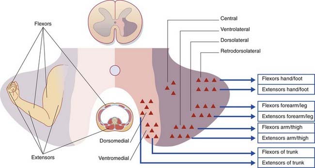
The segmental levels of the six somatomotor cell columns are listed in Table 16.1. The autonomic nervous system is represented by the intermediolateral cell column.
Table 16.1 The somatomotor cell columns
| Cell column | Muscles |
|---|---|
| Ventromedial (all segments) | Erector spinae |
| Dorsomedial (T1–L2) | Intercostals, abdominals |
| Ventrolateral (C5–C8, L2–S2) | Arm/thigh |
| Dorsolateral (C6–C8, L3–S3) | Forearm/leg |
| Retrodorsolateral (C8, T1, S1–S2) | Hand/foot |
| Central (C3–C5) | Diaphragm |
Cell types
Renshaw cells
The axons of the α motor neurons give off recurrent branches which form excitatory, cholinergic synapses upon inhibitory internuncial neurons called Renshaw cells in the medial part of the anterior horn. The Renshaw cells form inhibitory, glycinergic synapses upon the α motor neurons. This is a classic example of negative feedback, or recurrent inhibition, through which the discharges of α motor neurons are self-limiting (cf. Clinical Panel 8.1).
Segmental-level inputs to α motor neurons
Segmental-level inputs to a flexor α motor neuron include the following:
Descending Motor Pathways
Important pathways descending to the spinal cord are the following:
Corticospinal tract
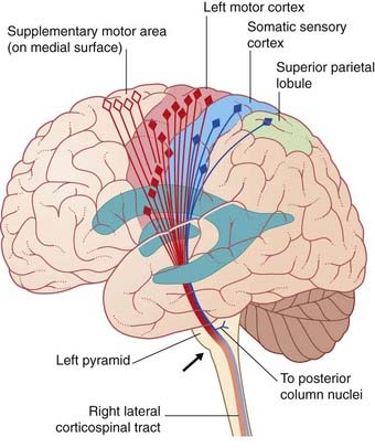
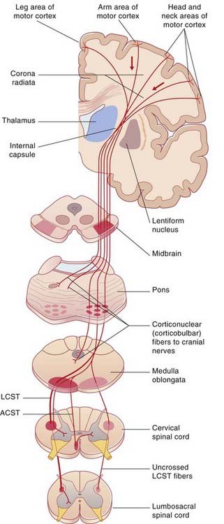
The corticospinal tract is the great voluntary motor pathway. About 40% of its fibers take their origin from the primary motor cortex in the precentral gyrus. Other sources include the supplementary motor area on the medial side of the hemisphere, the premotor cortex on the lateral side, the somatic sensory cortex, the parietal lobe, and the cingulate gyrus (Figure 16.2). The contributions from the two sensory areas mentioned terminate in sensory nuclei of the brainstem and spinal cord, where they modulate sensory transmission.
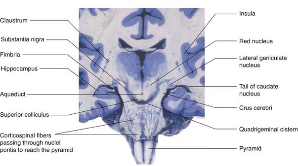
The corticospinal tract descends through the corona radiata and posterior limb of the internal capsule to reach the brainstem. It continues through the crus of the midbrain and the basilar pons to reach the medulla oblongata (Figure 16.3). Here it forms the pyramid (hence the synonym, pyramidal tract).
During its descent through the brainstem, the corticospinal tract gives off fibers which activate motor cranial nerve nuclei, notably those serving the muscles of the face, jaw, and tongue. These fibers are called corticonuclear (Figure 16.4). (The term ‘corticobulbar’ is sometimes used, but ‘bulb’ is open to different interpretations.)
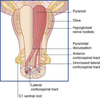
Just above the spinomedullary junction (Figure 16.5):
The corticospinal tract contains about one million nerve fibers. The average conduction velocity is 60 m/s, indicating an average fiber diameter of 10 µm (‘rule of six’ in Ch. 6). About 3% of the fibers are extra large (up to 20 µm); they arise from giant neurons (cells of Betz), located mainly in the leg area of the motor cortex (Ch. 26). All corticospinal fibers are excitatory and appear to use glutamate as their transmitter substance.
Targets of the lateral corticospinal tract
Distal limb motor neurons
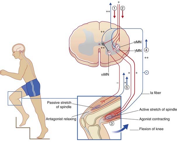
As mentioned already in Chapter 10, the α and γ motor neurons are coactivated by the LCST during a given movement, so that spindles in the prime movers are signaling active stretch while those in the antagonists are signaling passive stretch.
Ia inhibitory internuncials
Also located in the intermediate gray matter are the Ia inhibitory internuncials, and these are the first neurons to be activated by the LCST during voluntary movements. Activity of the Ia internuncials causes the antagonist muscles to relax before the prime movers (agonists) contract. In addition, it renders the antagonists’ motor neurons refractory to stimulation by spindle afferents passively stretched by the movement. The sequence of events is shown in Figure 16.6 and its caption for voluntary flexion of the knee.