Case
Age
Sex
Diagnosis
Recipient
Graft cut flow (ml/min)
1
44
F
R IC blood blister aneurysm
MCA (M2)
50
2
53
F
L IC blood blister aneurysm
MCA (M2)
20
3
68
F
R IC occlusion
MCA (M3)
30
4
65
M
BA trunk giant aneurysm
PCA (P3)
40
5
46
M
R PCA (P1) aneurysm
PCA (P2)
100
6
30
M
L PCA (P1/2) large aneurysm
PCA (P2)
30
7
77
M
L PCA (P1/2) giant aneurysm
PCA (P2)
30
Operative Technique
The surgical technique used was as follows: An STA main trunk was dissected in the length of 2 cm, and a 10–15 cm saphenous vein was harvested from the medial ankle of the lower thigh. A fronto-temporal craniotomy was made for the approach to the recipient artery and lesion. The recipient artery (M2 of the MCA or P2/3 of the PCA) was exposed through a trans-Sylvian approach. End-to-end anastomosis between the STA main trunk and the vein graft was made using interrupted sutures with 9-0 nylon sutures. A discrepancy in the size between the vein graft (generally 2–2.5 mm in diameter) and the STA main trunk (1.5–2 mm in diameter) can be compensated for using beveled end-to-end anastomosis with a fish-mouthed cut of the STA main trunk. The cut flow of the graft was measured. The end-to-side anastomosis between the short interposition vein graft (Ca 10 cm to the MCA and 15 cm to the PCA) and the intracranial recipient artery was then performed using interrupted sutures or running sutures with 10-0 nylon sutures. After confirming the patency of the bypass, an approach to the lesion was undertaken.
Results
Successful bypass surgery using short interposition vein grafts was therefore achieved. In six of seven patients, the perioperative course was uneventful. One patient with a ruptured right PCA (P1) aneurysm died of initial damage from a severe subarachnoid hemorrhage. All of the bypasses were patent. Intraoperative flow measurements confirmed a moderate flow-carrying capacity of the STA main trunk-interposition short vein grafts (20–100 ml; mean 43 ml/min).
Illustrative Case
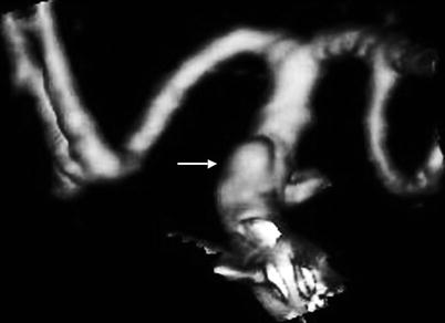
Case 2: A 53-year-old female patient presented with a WFNS grade 3 subarachnoid hemorrhage. CT demonstrated a diffuse subarachnoid hemorrhage and 3D CT angiography demonstrated a so-called blood blister-like aneurysm of the left internal carotid artery (Fig. 1). An STA main trunk to the left M2 bypass using interposition short vein graft was undertaken (Fig. 2a, b), and the aneurysm was treated using a wrap and clip technique (Fig. 2b). Intraoperative flow measurements of the cut flow of the bypass graft confirmed a rate of 30 ml/min. The patient tolerated the operation well. Figure 3 demonstrates the operative scar on the left medial ankle of the lower thigh from which the saphenous vein was harvested. Post-operative 3D CTA demonstrated good patency of the bypass graft (Fig. 4). The patient’s post-operative course was uneventful, and the outcome was scaled as a good recovery according to the Glasgow Outcome Score.
 of Combined Coiling and Neuroendoscopy in the Treatment of Intraventricular Hemorrhage Due to Ruptured Aneurysm
 Endarterectomy for Pseudo-occlusion of the Cervical Internal Carotid Artery
 Mini Supra-orbital Approach for Cerebral Aneurysm of the Anterior Portion of the Circle of Willis
 Exclusion of Unruptured Middle Cerebral Artery Aneurysms: Experience of 126 Cases
 Bypass Technique for Contemporary Revascularization of Unilateral MCA and Bilateral Frontal Territories in Moyamoya Vasculopathy
 of Plaque Location Using Indocyanine Green Videoangiography During Carotid Endarterectomy


Related posts:
of Combined Coiling and Neuroendoscopy in the Treatment of Intraventricular Hemorrhage Due to Ruptured Aneurysm
Endarterectomy for Pseudo-occlusion of the Cervical Internal Carotid Artery
Mini Supra-orbital Approach for Cerebral Aneurysm of the Anterior Portion of the Circle of Willis
Exclusion of Unruptured Middle Cerebral Artery Aneurysms: Experience of 126 Cases
Bypass Technique for Contemporary Revascularization of Unilateral MCA and Bilateral Frontal Territories in Moyamoya Vasculopathy
of Plaque Location Using Indocyanine Green Videoangiography During Carotid Endarterectomy
Stay updated, free articles. Join our Telegram channel
Full access? Get Clinical Tree
