42
Cavernous Sinus Anatomy
Jaime Gasco and Remi Nader
What is the structure of the cavernous sinus?
It is a hexahedron space at each side of the sella. It contains venous plexus, and the parasellar segment of the carotid artery along with cranial nerve VI
III, IV, V1, and V2 form part of the lateral wall.
There is a double dural layer in the posterior and lateral aspect of the cavernous sinus.
What are its borders?
Anterior: superior orbital fissure
Medial: sella and both clinoids
Posterior: petroclival ligament/tentorium
Posteromedial: clivus
Inferolateral: funnel-shaped area around foramen lacerum
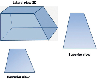
Fig. 42.1 Three-dimensional schematic representation of the cavernous sinus with superior and posterior views.
< div class='tao-gold-member'>


