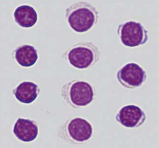
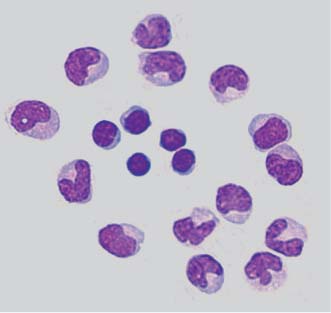
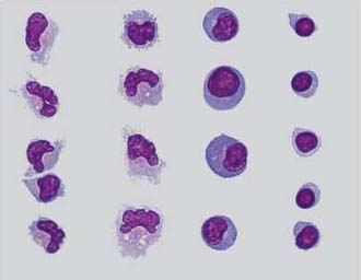
2 Cell Populations in the Normal Cerebrospinal Fluid H. Kluge, E. Linke, V. Wieczorek, K. Zimmermann, H.-J. Kuehn The normal lumbar cerebrospinal fluid (CSF) obtained by an uncomplicated lumbar puncture has a cell count of up to 5/μL, consisting exclusively of lymphocytes and monocytes in a ratio of approximately 70:30 (in sediment obtained by centrifugation) or 60:40 (after spontaneous sedimentation in a Sayk chamber, cf. Lymphocytes and Monocytes below). Rarely, normal CSF obtained in this way will contain cells or cell clusters belonging to the structures that enclose the CSF space. These cells, designated as surface epithelium in the consensus reporting form in Figure 1.1 (see also The CSF Cytology Report, Chapter 1 and Cells and Cell Clusters from the Structures Enclosing the CSF Space below), are more commonly seen after complicated lumbar punctures and in ventricular CSF. Their presence in lumbar CSF is to be considered a secondary effect of manipulation, i.e., as a CSF artifact. Further artifactual findings include bone marrow cells obtained by unintended aspiration of the marrow space as well as skin and cartilage cells (see Lumbar Puncture Artifacts below). Possible secondary artifacts due to contamination of the specimen after lumbar puncture (bacteria, fungi, dust particles, etc.) are not discussed in this chapter. The possibly artifactual admixture of blood in the CSF is discussed in Chapter 4. The lymphocytes in the cellular sediment of normal CSF stained with the May–Grünwald–Giemsa method are nonactivated, small isomorphic cells with a diameter of 5–8 μm. The round or mildly oval, turbid or coarsely granulated nucleus is surrounded by a very thin rim of cytoplasm, which is either practically indiscernible (such lymphocytes are said to have “naked nuclei”) or just wide enough to be visible. The cytoplasm is clear or, at most, mildly basophilic (Fig. 2.1). Immunocytochemical methods of cell differentiation (flow cytometry) reveal that about 93% of lymphocytes in normal CSF are T lymphocytes and about 1% are B lymphocytes. The distinction between these two types of cell is important in the differential diagnosis of inflammatory and infectious diseases. Venous blood, in contrast, contains 60–80% T lymphocytes and 10–30% B lymphocytes. Activated lymphocytes (activated B lymphocytes; many earlier authors classified these cells according to their cytoplasmic size and basophilia as a highly heterogeneous category of lymphoid cells, see Chapter 3, Cytological Findings in Infectious and Inflammatory Diseases) are rarely present in normal CSF, and their final differentiated type—the plasma cell—is never found in it. These cell types and the question of the origin of normal and activated lymphocytes in CSF are discussed in Chapter 3. The monocytes in the cellular sediment of normal CSF stained with the May–Grünwald–Giemsa method are of hematogenic (monocytopoietic) origin. Before passing into the CSF, however, they are present as “free” cells in the extracellular spaces of the meninges (including the stroma of the choroid plexus). Here, under pathological conditions, they may become transformed into activated monocytes and then into macrophages (see Chapter 4). As these monocytes are surrounded by a quite different milieu than blood, and because they have to migrate twice to find their way into the CSF (first across the vascular endothelium and then through the epithelial/ependymal cell layers), “normal” CSF monocytes have a highly varied morphology; at times, they almost resemble activated monocytes. With diameters of 15–20 μm, they are about four times as large as the normal CSF lymphocytes. The peripherally located nucleus is oval, or kidney- or horseshoe-shaped, or lobulated, and sometimes contains pale nucleoli. The cytoplasm is smoky gray or, at most, mildly basophilic near the cell membrane. Any basophilic coloring of the cytoplasm, or the occasional appearance of a few vesicular structures within it, indicates an incipient activated state. Figure 2.2 shows diverse structural variants of “normal” CSF monocytes surrounding five normal lymphocytes in the center of the figure. Although this chapter is principally devoted to normal cell morphology, we have included some normal forms and contrasting activated forms of lymphocytes and monocytes in Figure 2.3 for didactic purposes. Note the differences between normal and activated types with respect to cell size and shape, nuclear size and shape, and above all, cytoplasmic structure and staining. Activated monocytes are sometimes considerably larger than the ones shown in Figure 2.3 (see the cytological illustrations in Chapters 3 and 4 for examples). Fig. 2.1 Nonactivated lymphocytes in normal CSF. Left column: lymphocytes with “naked nuclei,” i.e., the cytoplasm is barely discernible as a clear or only slightly colored rim around the nucleus. Middle column: lymphocytes with a significantly larger amount of clear cytoplasm. Right column: lymphocytes with a thin, mildly basophilic rim of cytoplasm (incipient activation). Fig. 2.2 Nonactivated monocytes in normal CSF (outer circle of cells), some of which show early signs of activation, and, for comparison, lymphocytes with a thin, mildly basophilic rim of cytoplasm (the five cells in the center).
Lymphocytes and Monocytes
Related posts:
 Introduction
 Pathological CSF Cell Findings in Cysts
 Pathological CSF Cell Findings in Infectious and Inflammatory Diseases of the Central Nervous System
 Pathological CSF Cell Findings in Intracranial Hemorrhage and Traumatic and Hypoxic-Ischemic Brain Injury
 Pathological CSF Cell Findings in Primary and Metastatic CNS Tumors, Malignant Lymphoma, and Leukemia
 Smell and Tast
Stay updated, free articles. Join our Telegram channel

Full access? Get Clinical Tree


