Description of Cervical Osteotomy Classification System
The cervical osteotomy classification system provides a common nomenclature to more effectively and objectively communicate the type of surgical resection(s) performed. It is not intended to provide indications for surgery or to assist in the determination of the appropriate surgical approaches. The system is a 7-point scale based on anatomical resections that are graded according to the potential for destabilization (▶ Table 8.1, ▶ Fig. 8.1). It is designed to be comprehensive as to include the wide range of surgical resections that may be performed for cervical deformity correction.
Osteotomy grade | Resection | Description | Surgical approach modifiers |
1 | Partial facet joint resection or anterior discectomy and partial uncovertebral joint resection | Anterior cervical discectomy and partial uncovertebral joint resection, or partial facet resection | A, P, AP, PA, APA, PAP |
2 | Complete facet joint resection | Resection of both superior and inferior facets at a given segment | P, AP, PA, APA, PAP |
3 | Partial or complete corpectomy | Partial or complete corpectomy including discs above and below | A, AP, PA, APA, PAP |
4 | Complete uncovertebral joint resection to transverse foramen | Anterior osteotomy through lateral vertebral body and uncovertebral joints into transverse foramen | A, AP, PA, APA, PAP |
5 | Opening wedge osteotomy | Complete posterior element resection with osteoclastic fracture and open wedge creation | P, AP, PA, APA, PAP |
6 | Closing wedge osteotomy | Complete posterior element resection and pedicle resection with closing wedge creation | P, AP, PA, APA, PAP |
7 | Vertebral column resection | Resection of one or more entire vertebral bodies and discs including complete uncovertebral joint and posterior lamina and facets | AP, PA, APA, PAP |
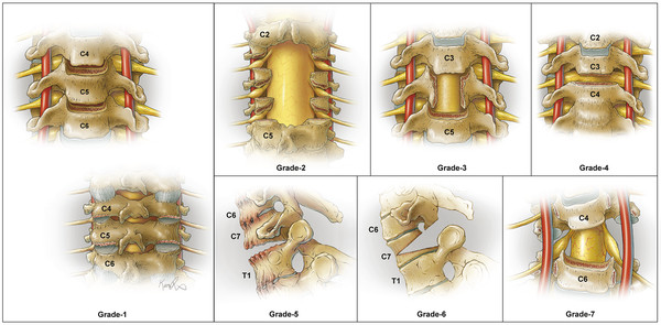
Fig. 8.1 Illustrations depicting the 7 cervical osteotomy types, which are based on anatomical resections according to progression of destabilization. Used with permission from Xavier Studio.
A given case may involve multiple resection types. The highest grade of osteotomy performed is designated as the major osteotomy, whereas other lower grades of osteotomies performed are designated as minor osteotomies. Additionally, modifiers are used to designate the surgical approach(es): anterior (A), posterior (P), anterior-posterior (AP), posterior-anterior (PA), anterior-posterior-anterior (APA), and posterior-anterior-posterior (PAP).
8.2.1 Grade 1: Partial Facet Joint Resection or Discectomy and Partial Uncovertebral Joint Resection
A Grade 1 osteotomy involves a partial joint resection. It may be performed via an anterior approach that includes a discectomy and partial uncovertebral joint resection, a posterior approach that includes partial resection of the facet joint, or through combinations of both approaches. A Grade 1 osteotomy has limited capacity for deformity correction, but it may be applied over multiple levels to cumulatively facilitate realignment. When performed posteriorly, it offers the potential to promote fusion by removing cartilage from the facet surfaces.
Grade 1 osteotomies may be performed through anterior or posterior approaches (modifiers A or P), or a combination of both approaches (AP, PA, APA, or PAP). Whether performed anteriorly or posteriorly, Grade 1 osteotomies require mobility of the opposite column (posterior or anterior, respectively) to achieve correction.
Case Example: Grade 1, Modifier A
A 51-year-old man presented with signs of cervical myelopathy and left upper extremity weakness and atrophy. Imaging demonstrated a loss of cervical lordosis with multilevel disc degeneration but no evidence of facet ankylosis (▶ Fig. 8.2). He underwent preoperative traction for 48 hours followed by a multilevel anterior cervical discectomy with partial uncovertebral joint resection and interbody fusion at C3–C4, C4–C5, C5–C6, and C6–C7.
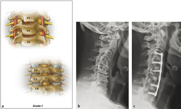
Fig. 8.2 (a) Grade 1 osteotomy, anterior cervical discectomy, partial uncovertebral joint resection, or partial facet joint resection. Pre- (b) and postoperative radiographs (c) depicting anterior cervical discectomies with partial uncovertebral joint resections at C3–C4, C4–C5, C5–C6, and C6–C7 (Grade 1, modifier A).
8.2.2 Grade 2: Complete Facet Resection
A Grade 2 osteotomy involves resection of both superior and inferior facets at a given segment. Other posterior elements (i.e., the lamina, ligamentum flavum, and spinous process) may be performed as part of a Grade 2 osteotomy, but this grade of resection does not include any portion of the vertebral body. This type of resection is analogous to those commonly performed for deformity correction in the thoracic and lumbar spine, which have several variations and terms, including Smith-Petersen, Ponte, polysegmental, chevron, and extension osteotomies among others. 15,16,17 Grade 2 osteotomies are often performed at multiple levels to facilitate a greater magnitude of deformity correction.
A posterior approach (modifier P) is used for Grade 2 osteotomies, but anterior column mobility is necessary. Thus, Grade 2 osteotomies may be performed in combination with anterior soft-tissue release procedures (modifiers AP, PA, APA, or PAP).
Case Example: Grade 2, Modifier P
A 64-year-old man presented with neck pain and difficulty maintaining horizontal gaze. Imaging demonstrated a loss of cervical lordosis (▶ Fig. 8.3) but no evidence of ankylosis of the anterior disc spaces (not shown). The deformity was considered to be partially reducible. A single-stage operation was performed consisting of posterior instrumentation and fusion from C2 to T10 and posterior column osteotomies with complete facet joint resections from C3–C4 to C7–T1.
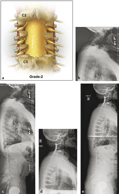
Fig. 8.3 (a) Grade 2 osteotomy, complete facet joint resection. Pre- (b,c) and postoperative radiographs (d,e) depicting osteotomies with complete facet joint resections at C3–C4, C4–C5, C5–C6, C6–C7, and C7–T1 and posterior instrumentation and fusion from C2 to T10 (Grade 2, modifier P).
8.2.3 Grade 3: Partial or Complete Corpectomy
A Grade 3 osteotomy involves partial or complete resection of a vertebral body, including the adjacent discs. In addition to providing anterior release, performing a corpectomy facilitates decompression of the spinal canal and foramina. A structural graft or expandable cage is then inserted into the corpectomy defect to enable restoration of cervical lordosis and provide arthrodesis. A ventral strategy provides an optimal approach for decompression of the spinal cord and enables placement of the interbody graft in a structurally favorable location for correction of the deformity.
An anterior approach (modifier A) is used for Grade 3 osteotomies, but mobility of the posterior column (i.e., facet joints) is necessary to maximize deformity correction. Thus, Grade 3 osteotomies may be performed in combination with posterior release procedures (modifiers AP, PA, APA, or PAP).
Case Example: Grade 3, Modifier A
A 72-year-old woman presented with neck pain and cervical myelopathy. Imaging demonstrated a Grade 3 spondylolisthesis of C3 in the setting of an autofusion across the disc space at C4 and C5 (▶ Fig. 8.4). A single-stage anterior procedure was performed with corpectomies at C4 and C5 (major osteotomy, Grade 3, modifier A), discectomy at C6–C7 (minor osteotomy, Grade 1, modifier A), placement of a fibular strut allograft between C3 and C6, placement of a bone allograft spacer between C5 and C6, and plating from C3 to C7.
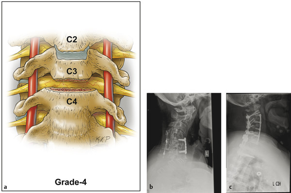
Fig. 8.4 (a) Grade 3 osteotomy, partial or complete corpectomy. Pre- (b) and postoperative radiographs (c) depicting complete anterior corpectomies at C4 and C5 (Grade 3, modifier A).
Related posts:
 Congenital Cervical Deformity and Hemivertebra
Relationship of Cervical Spondylotic Myelopathy to Cervical Deformity
Medical Complications
Cervical Pedicle Subtraction Osteotomy for Correction of Sagittal Deformities
C1-C2 Joint Osteotomy and Reduction of Vertical Deformity
Cervical Opening Wedge Osteotomy
Congenital Cervical Deformity and Hemivertebra
Relationship of Cervical Spondylotic Myelopathy to Cervical Deformity
Medical Complications
Cervical Pedicle Subtraction Osteotomy for Correction of Sagittal Deformities
C1-C2 Joint Osteotomy and Reduction of Vertical Deformity
Cervical Opening Wedge Osteotomy
Stay updated, free articles. Join our Telegram channel

Full access? Get Clinical Tree


