Complex Regional Pain Syndrome


Complex Regional Pain Syndrome


Jasvinder Chawla

Venkata Reddy


The International Association for the Study of Pain (IASP) suggests the terms complex regional pain syndrome type I (CRPS type I) for reflex sympathetic dystrophy and CRPS type II for causalgia. The sole differentiating criterion between CRPS type I and type II is the presence of a known nerve injury in CRPS type II. CRPS type I usually follows an initiating noxious event, is not limited to the distribution of a single nerve, and is apparently disproportionate to the inciting event. At some point, there is associated edema, changes in skin blood flow, abnormal sudomotor activity in the region of the pain, allodynia, or hyperalgesia. CRPS type I is associated with a variety of precipitating factors (
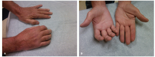
Table 51.1); trauma is the most common precipitating event (Fig. 51.1A and B).


I. PATHOPHYSIOLOGY

Various hypotheses include peripheral mechanisms, central mechanisms, or psychogenic factors.

A. Peripheral mechanisms do not address the spread of pain beyond a dermatomal terri tory and pain occurring in patients without nerve injury. They are of four types.

1. After an inciting event, a subset of C-polymodal nociceptors develop sensitivity to sympathetic stimulation and thus may be stimulated by noradrenalin. An alternative explanation is that noradrenalin may act indirectly through the release of prostaglandins that stimulate the nociceptors.

2. Sympathetic efferents cause abnormal activation of peripheral nociceptors.

3. Aberrant nerve sprouts generated at the site of injury develop into neuromas.

4. Artificial synapses form at the site of nerve injury and allow “ephaptic” transmission between sympathetic efferent and sensory afferent fibers.

B. Central mechanisms are of two types.

1. Self-sustaining loops of abnormal interneuronal firing in the dorsal horn, after being propagated by a peripheral irritative focus, give rise to ascending projections of pain and descending sympathetic hyperactivity.

2. Long-term sensitization or “wind-up” of wide-dynamic-range neurons in the spinal cord resulting from ongoing nociceptive stimulation from the periphery. The sensitized wide-dynamic-range neurons then respond to activity in large diameter A-mechanoreceptors that are activated by light touch. The pain threshold is reduced and previously subthreshold stimuli are then perceived as painful.

C. Psychogenic factors. A major issue in the diagnosis and management of CRPS is the lack of properly controlled comparison studies of placebo with sympathetic blockade. Furthermore, a fair number of patients with neuropathic pain improve with injec tions of placebo. In some patients, symptoms may be conversion-somatization of an underlying psychiatric condition, because these patients seem to respond to cognitive psychotherapy.


II. DIAGNOSIS AND COURSE


A. Diagnosis.


1. IASP diagnostic criteria include the following:


  • Presence of an initiating noxious event or a cause of immobilization.


  • Persistent pain, allodynia, or hyperalgesia, with ongoing pain disproportionate to the inciting event.


  • Evidence at some time of edema, changes in skin blood flow, or abnormal sudomotor activity in the region of the pain.


  • Diagnosis excluded by the existence of conditions that would otherwise account for the degree of pain and dysfunction.


FIGURE 51.1 A: and B: A 59-year old man was drilling a hole in concrete when the drill kicked back and jerked his hand. Patient did not seek medical care for 2 weeks. When he did seek medical care, he found he had crushed two wrist bones, the trapezium, and triquetral. The patient was casted for 4 weeks and then developed severe pain and soreness of the right hand. On examination the dorsum of the right hand was purplish-reddish and very shiny, and there was more hair on the right hand than on the left. (Courtesy of Dr. Jose Biller.)


TABLE 51.1 Precipitating Factors in the Development of CRPS Type I


Soft-Tissue Injury


Malignancy


Fracture


Arthritis


Sprain


Bursitis


Joint dislocation


Peripheral nerve injury


Operative procedures


Carpal tunnel release


Immobilization with a cast or splint


Venipuncture


Arthroscopic surgery


Myocardial infarction


Brachial plexopathy


Polymyalgia rheumatica


Radiculopathy


Myelopathy


Stroke


Dental extraction


Spinal cord injury


Prolonged bed rest


Drugs (isoniazid, phenobarbital, ergotamine and cyclosporine)


Criteria (b) through (d) must be satisfied.


2. Modified diagnostic criteria.

A study validated the IASP diagnostic criteria but showed that the CRPS criteria have inadequate specificity and are likely to lead to overdiagnosis. The investigators proposed the following modified research diagnostic criteria for CRPS:


  • Continuing pain disproportionate to any inciting event.


  • At least one symptom in each of the four following categories:


    • Sensory: hyperesthesia.


    • Vasomotor: temperature asymmetry, skin color changes, skin color asymmetry, or a combination of these signs.


    • Sudomotor edema: edema, sweating changes, sweating asymmetry, or combination of these symptoms.


    • Motor/trophic: decreased range of motion, motor dysfunction (weakness, tremor, and dystonia), trophic changes (hair, nails, and skin), or a combination of these symptoms.


  • At least one sign in two or more of the following categories:


    • Sensory: evidence of hyperalgesia to pin prick or allodynia to light touch.


    • Vasomotor: evidence of temperature asymmetry, skin color changes, asymmetry, or a combination of these signs.


    • Sudomotor/edema: evidence of edema, sweating changes, sweating asymmetry, or a combination of these symptoms.


    • Motor/trophic: evidence of decreased range of motion, motor dysfunction (weakness, tremor, and dystonia), trophic changes (hair, nails, and skin), or a combination of these symptoms.

      These research criteria may help prevent overdiagnosis of CRPS and may improve the ability to differentiate CRPS from other types of neuropathic pain.


3. Diagnosis of CRPS type I is generally made on the basis of history and clinical find ings. Laboratory testing is utilized to exclude other diagnosis. Clinical features are summarized in Table 51.2. CRPS in children is often under recognized by physicians resulting in diagnostic delays. The lower extremities are more commonly affected than the upper extremities. Minor trauma remains the most frequent cause. The female pre ponderance is much greater among children than among adults; patients are typically pubertal adolescent girls.

A therapeutic response to sympathetic neural blockade should be carefully evaluated to identify responders to this diagnostic procedure. The pain-relieving effect should outlast the expected 6- to 12-hour effect of the local anesthetic.


4. Differential diagnosis.

CRPS type II (causalgia), unrecognized local lesions (e.g., fracture, strain, and sprain), traumatic vasospasm, cellulitis, Raynaud’s disease, thromboangiitis obliterans, arterial/venous thrombosis, diabetic painful neuropathy, radicular syndromes, and gout.


5. Methods

used to aid in difficult-to-diagnose cases of and various severities of CRPS type I include radiography and scintigraphy. Most laboratory tests for CRPS are based on detection of asymmetric vascular or sudomotor function between the affected and contralateral unaffected side.


  • Radiography. Plain radiographs may show patchy osteopenia in one half of all patients. Plain radiography remains useful in detecting or excluding other bony abnormalities.


  • Scintigraphy. A three-phase technetium bone scan (TPBS) is helpful in confirm ing the diagnosis and staging of CRPS type I. The sensitivity and specificity ranges from 54% to 100% and 85% to 98%, respectively. Given a variety of presentations of CRPS, a TPBS not only confirms the diagnosis, but it also helps exclude other diagnoses such as degenerative arthritis, benign or malignant bony lesions, or even metabolic bone diseases such as Paget’s disease, osteomyelitis, stress fracture, bone infarction, Reiter’s disease, and thoracic outlet syndrome, particularly if utilized in combination with SPECT/CT. A TPBS is used in three ways.


    • Blood flow phase. Rapid-sequence images of the involved extremity are obtained after intravenous (IV) injection of a radionuclide tracer to evaluate the vascularity of a region.


    • Blood pool phase. Images are obtained immediately after the blood flow phase to evaluate regional perfusion, including that of soft tissue.


    • Bone scan phase. Static images are obtained 2 to 3 hours after initial injection to detect abnormal osteoblastic activity, reflected locally as increased periarticular uptake in the affected extremity.


  • Quantitative sudomotor axon reflex testing (QSART). Resting sweat output and stimulated sweat output as measured by QSART can help detect sudomotor asymmetry between the affected and contralateral unaffected side. Thermography and infrared thermometry are also used to measure skin temperature as an index of blood flow.


TABLE 51.2 Clinical Features of CRPS Type I


Feature


Example


Autonomic deregulation


Temperature, vasomotor, and sudomotor instability


Blood flow alterations


Hyperhydrosis, hypohydrosis, edema, and discoloration


Sensory abnormalities


Hyperalgesia burning pain, hyperpathia, allodynia, dysesthesia and hemisensory impairment


Motor dysfunction


Weakness, tremor, and dystonia


Trophic changes


Skin thinning, hair loss, brittle nails, and changes in structure of both superficial and deep tissues


Psychological disturbances


Anxiety, depression, and suicidal ideation


Radiologic changes


Patchy osteoporosis, soft-tissue edema, and articular erosion


B. Course.

CRPS type I can progress through three stages (Fig. 51.2).


1. Stage 1 (acute).

Pain is described as aching or burning, aggravated by physical contact or emotional upset, and typically restricted to a vascular or peripheral nerve or root territory. Some patients report paresthesias or burning distal pain. Hyperalgesia may be present, as may allodynia to light touch, thermal stimulation, deep pressure, or joint movement. Tissue swelling and local vascular, bony, and trophic changes occur in the affected part. Radiography may show diffuse bony changes. TPBS may show increased radionuclide uptake in all phases. Stage 1 usually occurs 1 to 3 months after injury.

Aug 18, 2016 | Posted by in NEUROLOGY | Comments Off on Complex Regional Pain Syndrome

Full access? Get Clinical Tree

Get Clinical Tree app for offline access