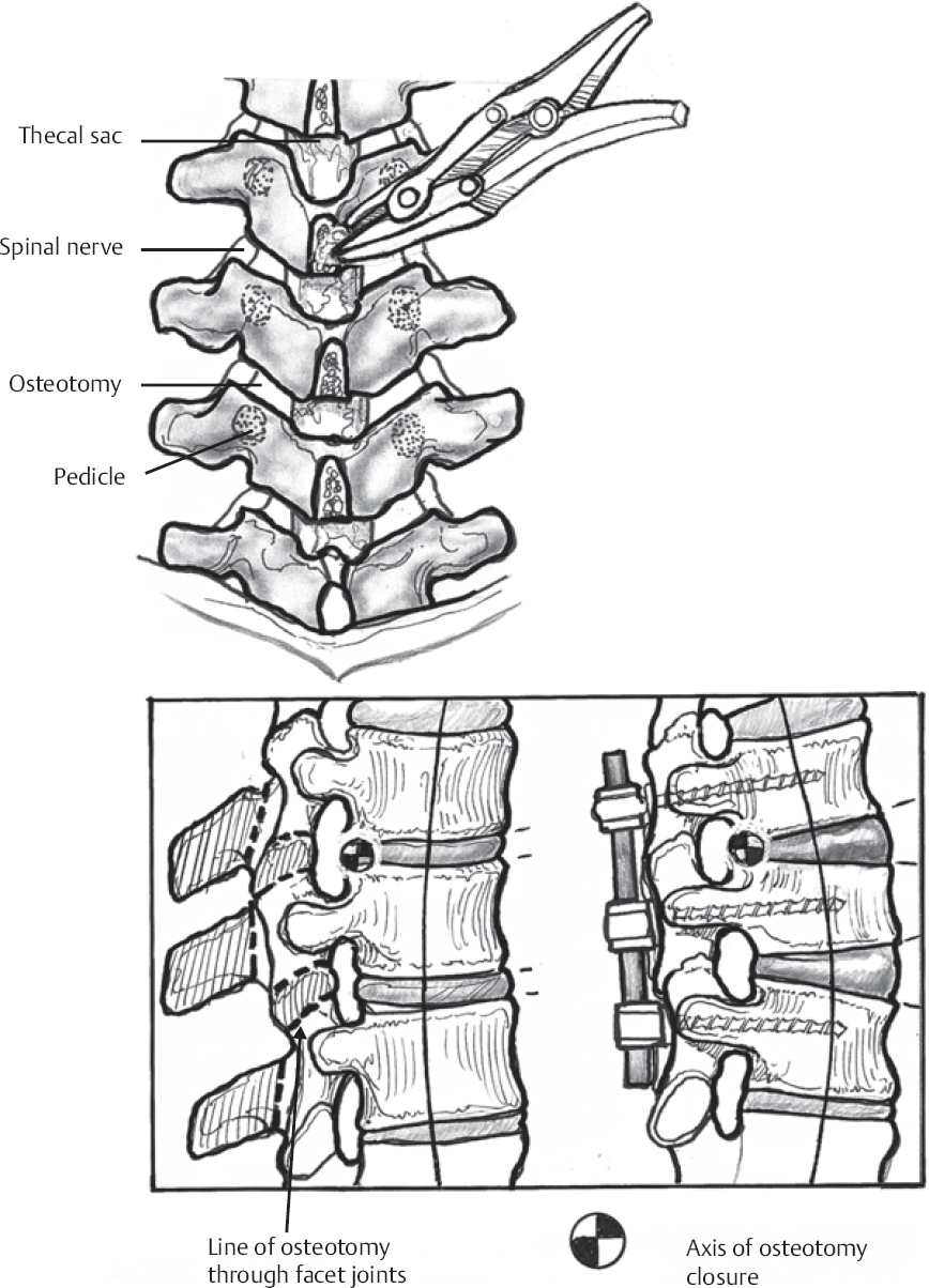
♦ Preoperative
Operative Planning
- Review imaging (plain radiographs, magnetic resonance imaging, myelogram/computed tomography myelogram)
Routine Equipment
- Basic lumbar laminectomy and fusion instruments
Special Equipment
- Thoracolumbar pedicle screw implants
- Iliac fixation system
- Neurophysiological monitoring for somatosensory evoked potentials, motor evoked potentials, and pedicle screw testing with triggered electromyography (EMG)
Operating Room Set-up
- Open-frame spinal table with traction set-up (optional)
- Headlight
- Two Bovie cauteries for simultaneous bilateral exposure
- Cell saver
- Smoke evacuator
Anesthetic Issues
- Sufficient intravenous access for blood transfusion
- Arterial line for blood pressure monitoring
- Wake-up test may be required during procedure
- For complex procedures, consider epsilon aminocaproic acid to reduce blood loss
♦ Intraoperative
Positioning
- Gardner-Wells tongs placed in standard position (1 cm above pinnae, inline with external auditory meatus)
- 15 lb of inline traction
- If using head holder instead of traction, ensure no ocular pressure
- Three pads on each side: 3 to 4 cm distal to axillae, proximal and distal to anterior superior iliac spine
- Hips extended to maximize lumbar lordosis
- Shoulders abducted and elbows flexed 90 degrees
- All pressure points well padded
Sterile Scrub and Prep
- As in posterior thoracic approach
Incision
- Linear incision extending from tip of spinous process one level proximal to upper instrumented vertebra (UIV) to spinous process of lower instrumented vertebra (LIV)
Exposure
- Bilateral subperiosteal exposure to tips of transverse processes of all levels to be included in construct
- Maintain integrity of supra- and interspinous ligament proximal to UIV and distal to LIV
- Avoid disruption of facet capsules of levels excluded from construct
- Thoroughly clean all soft tissue from dorsal bony surfaces of spine
Decompression
- Perform decompression (laminotomy, laminectomy, foraminotomy) at appropriate levels
Pedicle Screw Fixation
- Place bilateral pedicle screws at each segment
- Begin distally and work proximally
- Ensure appropriate screw length based on preoperative measurements and intraoperative confirmation
- Optimal SI screws are bi- or tri-cortical (exit ventrally at sacral promontory)
- Obtain anteroposterior (AP) and lateral radiographs to confirm proper screw placement
- Test triggered EMG threshold for each screw distal to T6
Iliac Fixation (Fig. 125.1)
- Used to prevent sacral screw pull out for long (greater than ~5-level) constructs crossing lumbosacral junction
- Thoroughly expose distal ilium, and remove soft tissue overlying sacroiliac joint (minimize disturbing joint itself).
< div class='tao-gold-member'>
Only gold members can continue reading. Log In or Register to continue
Stay updated, free articles. Join our Telegram channel

Full access? Get Clinical Tree


