Cranial nerves and cranial nerve nuclei
There are 12, bilaterally paired, cranial nerves (I–XII). These carry afferent and efferent fibres between the brain and peripheral structures, principally of the head and neck. The cranial nerves are individually named and numbered (Roman numerals, see below) according to the rostrocaudal sequence in which they attach to the brain (Fig. 10.1 and Table 10.1):
II optic
III oculomotor
IV trochlear
VI abducens
VII facial
VIII vestibulocochlear
X vagus
XI accessory
XII hypoglossal
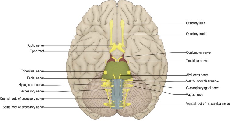
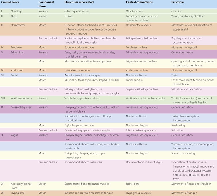
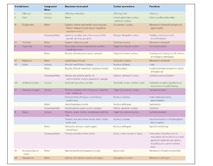
Table 10.1
Summary of components, connections and functions of the cranial nerves
The components are colour-coded according to their embryological origin (see also Fig. 1.11 and Fig. 10.2).
 |
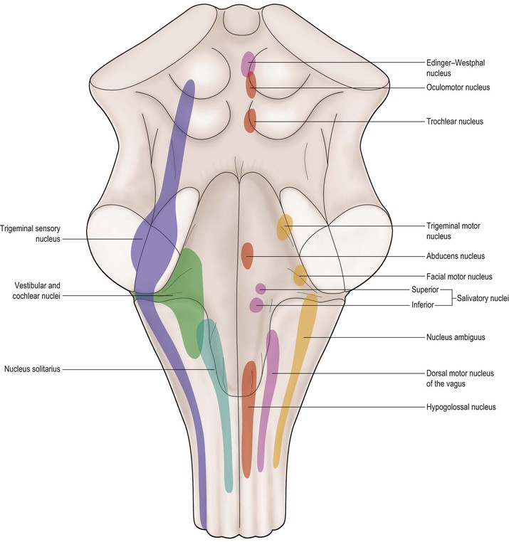
The first two cranial nerves attach directly to the forebrain, while the rest attach to the brainstem. The olfactory system is closely associated, both structurally and functionally, with parts of the forebrain collectively referred to as the limbic system; these, including cranial nerve I, are considered together in Chapter 16. The visual system and cranial nerve II are described in Chapter 15. Cranial nerves III–XII are associated with various nuclei within the brainstem, called the cranial nerve nuclei, which either receive cranial nerve afferents or contain the cell bodies of efferent neurones that have axons leaving the brain in cranial nerves. The locations of these nuclei are illustrated schematically in Figure 10.2. Some can readily be seen in stained sections of the brainstem (Figs 9.5–9.13).
Cranial nerve nuclei
Afferent nuclei
Fibres carrying general sensory information (touch, pressure, pain, temperature) from the head enter the brain through the trigeminal nerve at the level of the pons and terminate in the trigeminal sensory nucleus. This is a large nucleus that runs the whole length of the brainstem and extends caudally into the cervical spinal cord. Fibres conveying the special senses of motion/positional sense and hearing run in the vestibulocochlear nerve. They terminate in the vestibular and cochlear nuclei, respectively, which are located in the medulla, in and near to the lateral part of the floor of the fourth ventricle (sometimes referred to as the vestibular area). Visceral afferents, including taste fibres, terminate in the nucleus solitarius of the medulla.
Efferent nuclei
On the basis of their embryological derivation, the efferent cranial nerve nuclei can be divided into three groups, each lying in a discontinuous longitudinal column.
Nuclei of the somatic efferent cell column
The somatic efferent cell column lies near to the midline and consists of the nuclei of the III, IV, VI and XII nerves. The oculomotor nucleus lies in the ventral apex of the periaqueductal grey of the midbrain at the level of the superior colliculus (Fig. 9.13). Its efferent fibres run in the oculomotor nerve to innervate the levator palpebrae superioris muscle and all of the extraocular muscles, except the superior oblique and lateral rectus. The trochlear nucleus also lies in the midbrain, at the ventral border of the periaqueductal grey, but at the level of the inferior colliculus (Fig. 9.12). Fibres leave in the trochlear nerve, to innervate the superior oblique muscle of the eye. The abducens nucleus is located in the caudal pons beneath the floor of the fourth ventricle (Fig. 9.8). Its efferents run in the abducens nerve and they innervate the lateral rectus muscle. In the medulla lies the hypoglossal nucleus (Fig. 9.7), which innervates the intrinsic and extrinsic muscles of the tongue via the hypoglossal nerve.
Nuclei of the branchiomotor cell column
The branchiomotor cell column innervates striated muscles derived from the branchial arches. In the tegmentum of the mid-pons is located the trigeminal motor nucleus, which supplies fibres to the trigeminal nerve and innervates the muscles of mastication, tensor tympani, tensor veli palatini, mylohyoid and the anterior belly of the digastric muscle. In the caudal pontine tegmentum lies the facial motor nucleus. This innervates the muscles of facial expression and the stapedius muscle via the facial nerve. Within the medulla lies the nucleus ambiguus. This long nucleus sends motor fibres in the glossopharyngeal, vagus and cranial part of the accessory nerve to innervate the muscles of the pharynx and larynx.
Nuclei of the parasympathetic cell column
The parasympathetic cell column consists of preganglionic parasympathetic neurones that send axons into the III, VII, IX and X cranial nerves. The most rostral cell group constitutes the Edinger–Westphal nucleus, which lies in the midbrain periaqueductal grey matter adjacent to the oculomotor nucleus (Fig. 9.13). Its axons leave in the oculomotor nerve and pass to the ciliary ganglion in the orbit, from which postganglionic fibres innervate the sphincter pupillae and ciliary muscles within the eye.
In the pontine tegmentum lie two cell groups, the superior and inferior salivatory nuclei. The superior salivatory nucleus supplies preganglionic fibres to the facial nerve that terminate in the pterygopalatine and submandibular ganglia. Postganglionic fibres from the pterygopalatine ganglion innervate the lacrimal gland and the nasal and oral mucous membranes. Those from the submandibular ganglion innervate the submandibular and sublingual salivary glands. The inferior salivatory nucleus sends preganglionic fibres into the glossopharyngeal nerve. These terminate in the otic ganglion, which in turn sends postganglionic axons to the parotid salivary gland.
The largest preganglionic parasympathetic cell group lies in the medulla and constitutes the dorsal motor nucleus of the vagus (Fig. 9.7). Its rostral portion lies immediately beneath the floor of the fourth ventricle, lateral to the hypoglossal nucleus. Fibres leave in the vagus nerve and are widely distributed to thoracic and abdominal viscera.
Cranial nerves
III: Oculomotor nerve
The oculomotor nerve carries the majority of somatic motor axons that innervate the extraocular muscles responsible for moving the eye. It also contains preganglionic parasympathetic neurones that, via the intermediary of the ciliary ganglion, control the smooth muscle within the eye.
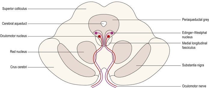
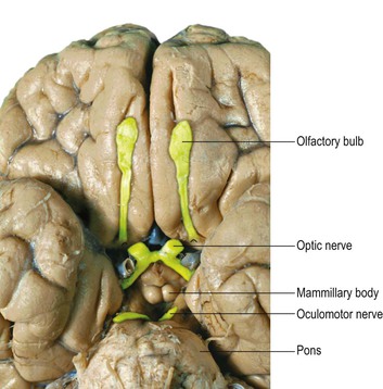
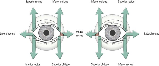
The motor neurones serving the extraocular muscles have their cell bodies in the oculomotor nucleus, which lies at the base of the periaqueductal grey of the midbrain at the level of the superior colliculus (Fig. 10.3). Preganglionic parasympathetic neurones arise from the nearby Edinger–Westphal nucleus (Fig. 10.3, see also Fig. 9.13). Fibres from both sources course ventrally through the midbrain tegmentum, many of them traversing the red nucleus, to exit on the medial aspect of the crus cerebri, within the interpeduncular fossa (Fig. 10.4). The oculomotor nerve passes between the posterior cerebral and superior cerebellar arteries (Fig. 7.2), then runs anteriorly, lying in the wall of the cavernous sinus (Fig. 7.11), before gaining access to the orbit through the superior orbital fissure. The oculomotor nerve supplies all of the extraocular muscles, with the exception of the superior oblique and lateral rectus and, thus, functions to elevate, depress and adduct the eyeball (Fig. 10.5). It also innervates the striated muscle of the levator palpebrae superioris, serving to elevate the upper eyelid. Preganglionic parasympathetic neurones from the Edinger–Westphal nucleus terminate in the ciliary ganglion, located within the orbit, behind the eyeball. From here, postganglionic neurones run in the short ciliary nerves to innervate the sphincter (constrictor) pupillae muscle of the iris and the ciliary muscle contained within the ciliary body.
Pupillary light reflex
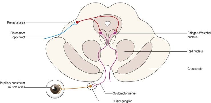
The amount of light entering the eye is regulated by the size of the pupil. Illumination of the retina causes constriction of the pupil through contraction of the sphincter pupillae muscle of the iris, thus reducing the amount of light reaching the retina. This is known as the direct light reflex (Fig. 10.6). Even if only one retina is illuminated (e.g. during clinical examination) the pupils of both eyes constrict. The constriction of the pupil of the non-illuminated eye is called the consensual light reflex. The afferent limb of the light reflex consists of a small contingent of optic tract fibres that pass directly from the eye to the pretectal area, just rostral to the superior colliculus, rather than to the lateral geniculate nucleus of the thalamus, where the majority of visual fibres terminate (see also Ch. 15). Neurones of the pretectal area project bilaterally to the Edinger–Westphal nuclei, from which efferent fibres leave in the oculomotor nerve.
Accommodation reflex
Fixation upon a nearby object, by convergence of the optic axes, involves concomitant contraction of the ciliary muscles to increase the convexity of the lens, thus focussing the image. It is also accompanied by pupillary constriction. The phenomenon involves the visual cortex, with corticobulbar fibres activating the parasympathetic neurones of the Edinger–Westphal nuclei bilaterally.
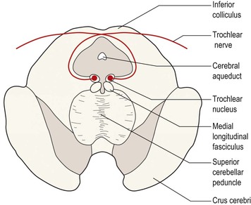
IV: Trochlear nerve
The thin trochlear nerve contains only somatic motor neurones. These arise in the trochlear nucleus, which lies in the ventral part of the midbrain periaqueductal grey, at the level of the inferior colliculus (Fig. 10.7). The efferent axons pass dorsally, around the periaqueductal grey, and decussate in the midline. The trochlear nerve emerges from the dorsal aspect of the brainstem (the only cranial nerve to do so) just caudal to the inferior colliculus (Fig. 10.8). The nerve courses round the cerebral peduncle to gain the ventral aspect of the brain (Fig. 10.9), passing between the posterior cerebral and superior cerebellar arteries (Fig. 7.2), as does the oculomotor nerve. It then runs anteriorly, lying in the lateral wall of the cavernous sinus (Fig. 7.11) and enters the orbit through the superior orbital fissure. It supplies just one muscle, the superior oblique. The action of the superior oblique is complex. Depending upon the starting position of the eyeball it can act to depress, abduct or intort the eye. Importantly, with the eye adducted, the superior oblique depresses the visual axis.
< div class='tao-gold-member'>
Related posts:


Stay updated, free articles. Join our Telegram channel

Full access? Get Clinical Tree


