Cranial Nerves
The Eye 2 – Fundi
BACKGROUND
The ophthalmoscope provides a light source and an optical system to allow examination of the fundus (Fig. 8.1).
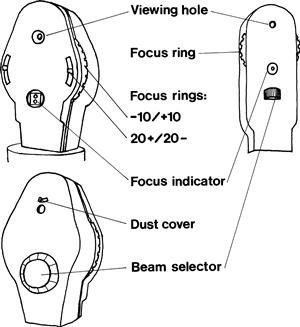
Its moving parts are:
The focus ring is used to correct (1) for your vision and (2) for the patient’s vision.
 TIP
TIP
An oblique view of the patient with his spectacles on tells you if he is long- or short-sighted and gives an idea of severity. If his face is smaller through his glasses, he is myopic; if his face is larger, he is hypermetropic. The degree indicates severity.
Beam selector choices are:
• narrow beam for looking at the macula
• target (like a rifle sight) to measure the optic cup
• green to look for haemorrhages (red appears as much darker).
WHAT TO DO
• Turn off the lights or draw the curtains.
• Check the focus is set at zero, and that the light works and is on the correct beam.
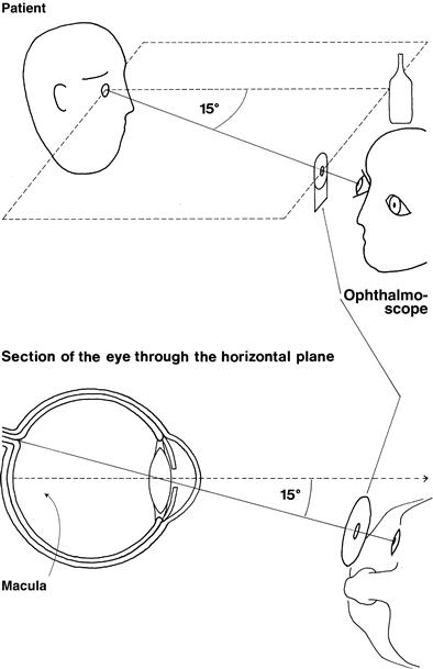
To examine the right eye (Fig. 8.2):
• Take the ophthalmoscope in your right hand.
• Approach the patient’s right side.
• The pupil should appear pink, as in bad flash photographs. This is the red reflex.
• Gradually move in towards the eye.
• Encourage the patient to keep looking at the distant point and not at the light.
• Bring the ophthalmoscope to within 1–2 cm of the eye.
• Keep the ophthalmoscope at the same level as the patient’s eye and the fixation point.
If the eye is approached as described, the optic disc should be in view. If it is not, focus on a blood vessel and follow it. The acute angles of the branches and convergence of artery and vein indicate the direction to follow. Alternatively, start again.
 TIP
TIP
It is essential to keep the patient’s eye, the point of fixation and the ophthalmoscope in the same plane.
To examine the left eye:
1 Look at the optic disc
Related posts:


Stay updated, free articles. Join our Telegram channel

Full access? Get Clinical Tree


