Chapter 151 Dorsal Thoracic and Lumbar Combined and Complex Techniques
Length of Construct
In adults, including the L5-S1 disc space in the fusion should be considered in any long construct that otherwise would have ended in the caudal lumbar spine. As outlined by Bridwell, indications for fusion to the sacrum in adults in a long construct include (1) L5-S1 spondylolisthesis, (2) previous L5-S1 laminectomy, (3) central or foraminal stenosis at L5-S1, (4) oblique takeoff of L5, and (5) “severe” degeneration of the disc.1 Although fusing to the sacrum does decrease a significant amount of motion, there is a significant risk of adjacent segment degeneration when the construct is stopped at L5 in the adult degenerative deformity population. A retrospective study by Edwards et al. found that in a population of patients who had undergone a thoracolumbar construct that ended caudally at L5 who preoperatively had a “healthy” L5-S1 disc, 61% of patients had developed advanced degenerative disease at this level over a mean follow-up of 5.6 years.2
Because of the risk of pseudarthrosis at the L5-S1 space, many authorities have advocated for the use of interbody support at this level. Polly et al. found that L5-S1 interbody support increases biomechanical stability, restores junctional lordosis, improves the lumbosacral fusion rate, and increases disc and foraminal height, thus decreasing foraminal stenosis.3 In the adult deformity patients undergoing spine fusion, the restoration or maintenance of sagittal balance has been found to have a significant impact on outcome.4 Restoring junctional lordosis at L5-S1 through placement of an interbody graft is a powerful technique to correct sagittal imbalance. This is due to the long moment arm that occurs when the L5-S1 disc space angulation is altered relative to the C7 vertebral body on which sagittal balance is based. To maximize the footprint of the graft as well as the restoration of lordosis, an anterior lumbar interbody fusion (ALIF) is a better procedure than a transforaminal lumbar interbody fusion (TLIF) at L5-S1.5 One must weigh the benefit of a larger interbody device against the morbidity of an anterior approach when selecting an anterior approach versus a TLIF or posterior lumbar interbody fusion (PLIF).
Long constructs that extend to the sacrum can be problematic from a fixation standpoint. Although the S1 pedicles are large, the sacrum is composed of primarily cancellous bone, resulting in a decreased pull-out strength compared with other pedicle screws. This has prompted many to attempt to place bicortical pedicle screws to capture the anterior and posterior cortical bone to increase the strength of the screw. Lehman et al. showed that the highest bone mineral density, and therefore the greatest insertional torque for pedicle screws, was in the anterior sacral promontory, so that from a biomechanical standpoint, the strongest S1 screws are the so-called “tricortical” screws. When screws are placed in this fashion, there is almost a 99% increase in the insertional torque.6 In addition to an L5-S1 interbody and the placement of tricortical S1 pedicle screws, iliac screws have been advocated as another method to offload the S1 pedicles to allow for a solid arthrodesis at L5-S1. Indications for iliac screws include constructs greater than three levels that end in the sacrum, revision surgery for L5-S1 pseudarthorsis, high-grade spondylolisthesis, or trauma or pathology that does not allow for adequate sacral fixation.7 Kuklo et al. evaluated 81 patients who underwent fusion procedures that included the L5-S1 space using S1 and iliac screws. Approximately 50% (n = 42) were for isthmic spondylolisthesis, whereas the remainder (n = 39) were for long constructs to the sacrum. The researchers found that even in patients who had previous iliac crest grafts taken, 94% (34/36) had iliac screws placed without screw loosening or iliac crest fracture. Overall, the fusion rate, to include revision surgeries, was 95.1%.8
Junctional Kyphosis
One of the reasons to plan and execute an appropriate-sized construct is to avoid proximal junctional kyphosis (PJK). In 1999, Lee et al. looked at 69 patients with adolescent idiopathic scoliosis (AIS) who underwent fusion up to T3. The investigators defined PJK as greater than 5 degrees above the summed normal of the angular segments from the proximal instrumented segment to T2. They found a 46% incidence of PJK. As would be expected, a predictor of postoperative PJK was preoperative kyphosis of more than 5 degrees above the proposed proximal instrumented level, indicating that these levels should be included in the construct to avoid this complication.9
In the adult deformity population, Kim et al. evaluated 161 patients with a minimum 5-year follow-up who had undergone long (more than five segments) dorsal constructs to determine the incidence and outcomes associated with PJK. The researchers found that at mean follow-up of 7.8 years, there was a 39% incidence of PJK defined either as a proximal junction sagittal Cobb angle of more than 10 degrees or as a proximal junction sagittal Cobb angle at least 10 degrees greater than the preoperative measurement. The time periods most notable for worsening of PJK were at 8 weeks postoperatively (59%) or after 2 years until final follow-up (35%). Scoliosis Research Society outcome measures did not show any significant difference in those with PJK as compared with those without except with self-image when PJK was more than 20 degrees. Age older than 55 years and combination anterior-posterior surgery were the only significant risk factors identified.10 The time course between the identification of PJK suggests two different populations, given that one group was relatively close to surgery, whereas the other was more remote. Further understanding of the similarities and differences of the early and late group may lead to changes in treatment strategies to better prevent PJK.
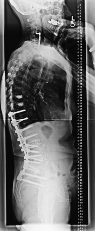
Another technique used to avoid PJK as well as strengthen the screw pull-out strength is vertebroplasty. Recently there has been some controversy over the effectiveness of this procedure in the setting of compression fractures, but the concept is applied differently in deformity surgery. At the proximal and sometimes distal screws, polymethylmethacrylate (PMMA) is injected under fluoroscopy into the screw tracks or through the screws themselves if the screw design allows it. Care is taken that there are no pedicle wall breaches allowing the egress of PMMA into the spinal canal or the production of embolic material. Once the PMMA is in place, typically 1 to 2 mL per side, then the pedicle screw is placed. Another scenario is to perform vertebroplasty at the level cephalad to the proximal instrumented vertebrae to prevent compression fractures leading to PJK (Fig. 151-1). Alternatively, this procedure can be performed in a postoperative setting. Preoperative PMMA augmentation of the planned proximal instrumented bodies is not recommended because it increases the difficulty of placement of the instrumentation.
In a cadaveric study designed to look at effects of cement augmentation of pedicle screws compared with extension with a flexible rod, Tan et al. found that in a corpectomy model, cement augmentation significantly reduced the range of motion and resulted in a more stable construct.11 In another cadaveric study, Becker et al. found that pedicle screws augmented with PMMA in an osteoporotic model had increased pull-out strength compared with non-PMMA augmented screws.12 In a cost-effectiveness analysis published in 2008, Hart et al. evaluated 28 women older than 60 years of age who had undergone fusion to the thoracolumbar region. Fifteen of these patients had undergone vertebroplasty in the adjacent level cranial to the proximal instrumented vertebrae. Proximal collapse occurred in none of the patients who had PMMA augmentation and in two (15.3%) patients who did not. Assuming a 15% decrease in the incidence of this problem, the researchers determined that the cost to prevent a single proximal junctional collapse was $46,240 using vertebroplasty, whereas the cost of revising the instrumentation in a patient with proximal junctional collapse was $77,432.13
Interbody Fusion
Interbody fusions are achieved by removing the intervertebral disc and placing fusion material such as autograft, allograft, or osteobiologics. Achieving fusion across the disc space has been shown to provide a biomechanically stiffer construct.3 But can interbody constructs stand alone? In 2006, Anjarwalla et al. evaluated their ALIF patients and divided them into four cohorts: stand-alone ALIF, ALIF with translaminar screws, ALIF with unilateral pedicle screws, and ALIF with bilateral pedicle screws. Using thin-section CT, the investigators reported a fusion rate of 51% in the stand-alone ALIF population, 58% in the ALIF population with translaminar screws, 89% in the ALIF population augmented by unilateral screws, and 88% fusion with ALIF augmented by bilateral pedicle screws.14 Some studies have advocated the use of percutaneous pedicle screws to augment ALIF with good clinical and radiographic results.15 Cadaveric studies have looked at the difference between stand-alone ALIF, ALIF with anterior plate, and ALIF with pedicle screws and found that augmentation of a stand-alone ALIF significantly reduced the range of motion and increased the stiffness of the construct. Overall, there was no consistent significant difference between an anterior plate and pedicle screws except that pedicle screws more effectively limited lateral bending.16,17
Related posts:
 Definition and Assessment of Dysfunctional Segmental Motion
Pathophysiology of Cervical Myelopathy: Biomechanics and Deformative Stress
Combined Ventral-Dorsal Surgery
Bone Void Fillers: Bone and Bone Substitutes
Medical Management of Neck and Low Back Pain
Posterior and Transforaminal Lumbar Interbody Fusion
Definition and Assessment of Dysfunctional Segmental Motion
Pathophysiology of Cervical Myelopathy: Biomechanics and Deformative Stress
Combined Ventral-Dorsal Surgery
Bone Void Fillers: Bone and Bone Substitutes
Medical Management of Neck and Low Back Pain
Posterior and Transforaminal Lumbar Interbody Fusion
Stay updated, free articles. Join our Telegram channel

Full access? Get Clinical Tree


