Dynamic MRI in the Seated Position Increases Insight into Diseases of the Lumbar Spine
Francis W. Smith
For the past 25 years, the lumbar spine has been studied using magnetic resonance imaging (MRI) in the supine position. This position is often the most comfortable for the patient, when in fact their symptoms are often worse when either standing or sitting. With the availability of an upright MRI scanner in which images can be made in the erect position, a number of studies have been performed to examine the changes in the appearances of both healthy and symptomatic patients in different postures. The effect of gravity on the body and the resultant differences in the appearances of the lumbar spine, abdomen, and pelvic contents are very obvious, as seen in Figure 5.1.
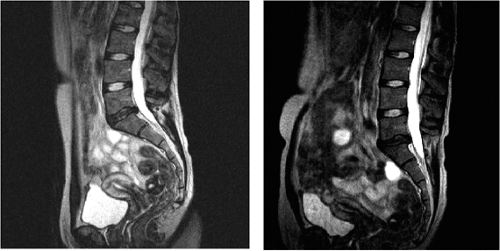 FIGURE 5.1 Supine/sitting erect. |
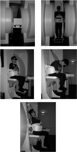 FIGURE 5.2 Upright MRI scanner. |
MRI Scanner
The MRI system used for these studies has been designed specifically to image patients in both the erect and supine positions. The system operates at 0.6 Tesla utilizing a resistive magnet with a horizontal field, transverse to the axis of the patient’s body (FONAR, Melville, NY). This provides a nonclaustrophobic open view from the magnet, allowing for unimpeded patient movement studies. A unique MRI-compatible, motorized patient handling system has been developed for the scanner, which allows for vertical (load bearing) to horizontal (supine) positioning of patients. Our system takes advantage of a 5-degree tilt in vertical studies to stabilize the patient and was used in all studies. The custom-built patient-handling system allows for patients to be imaged standing, sitting, lying supine, prone, or on their side, at any angle between vertical and 20 degrees in the Trendelenburg position. Because of its “open” design, patients are able to move within the scanner enabling images to be made in flexion and extension, in addition to the normal neutral position. A seat attachment has been developed to allow imaging in the sitting position. Data are acquired using a flexible, solenoidal radiofrequency (RF) receiver coil (Fig. 5.2).
Studies
The initial work with upright scanner concentrated on measuring the variations in the dimensions of the normal spinal canal, nerve root canals, and intervertebral discs. In one study, 29 male volunteers with no symptoms of low back pain, age 21 to 61 years (mean 32 years), were studied. The following observations were made, confirming the accepted knowledge as measured in cadaver studies:
We have shown a significant posture-dependent difference of the cross-sectional dural sac area at the intervertebral disc level in asymptomatic volunteers.
When changing posture from a supine to a standing position, the cross-sectional area of the dural sac increases (Fig. 5.3).
The smallest cross-sectional dural sac area was found in the supine position.
The dural sac contour took the appearance of a longitudinal ellipse in flexion and a transverse ellipse in extension and that cross-sectional dural sac area did not change significantly at the L3-4 and L4-5 disc level (Fig. 5.4) (1).
There are posture-dependent changes in the size and shape of the nerve root exit foramina (Figs. 5.5 5.6 5.7).Related posts:
 Adjacent Level Disc Biomechanics
 What is the Evidence Base for IDET?
 Total Lumbar Disc Arthroplasty: Overview of Clinical Results for Existing Implants
 Functional Lumbar Artificial Nucleus Replacement—DASCOR
 A New Approach to Lumbar Disc Prosthesis
 Posterior Nonfusion Stabilization of the Degenerated Lumbar Spine with Cosmic

Stay updated, free articles. Join our Telegram channel

Full access? Get Clinical Tree


