30 Electroencephalography
The EEG is an important neurological tool, for three main reasons:
We suggest you review the amino acid transmitters in Chapter 8 before reading about drug therapy in the Clinical Panels.
Neurophysiological Basis of the EEG
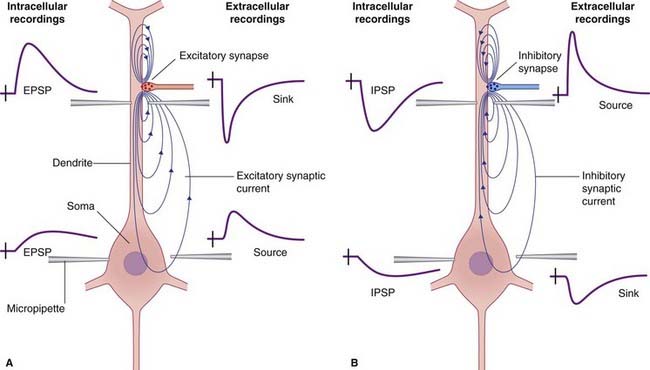
When small metallic disc electrodes are placed on the surface of the scalp, oscillating currents of 20–100 µV can be detected and are referred to as an electroencephalogram (EEG). Their origin is a direct consequence of the additive effect of groups of cortical pyramidal neurons being arranged in radial (outward-directed) columns. The columns relevant here are those beneath the surface of the cortical gyri. As the membrane potentials of these columns fluctuate, an electrical dipole (adjacent areas of opposite charge) develops. The dipole results in an electrical field potential as current flows through the adjacent extracellular space as well as intracellularly through the neurons (Figure 30.1). It is the extracellular component of this current that is recorded in the EEG and variations in both the strength and density of the current loops result in its characteristic sinusoidal waveform.
Technique
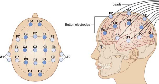
After careful preparation of the skin of the scalp to ensure good contact, electrodes are affixed in a placement that is in conformity with the 10–20 International System of Electrode Placement, when the scalp is divided into a grid in accordance with Figure 30.2.
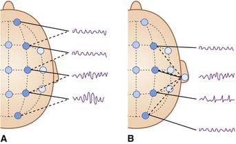
If varying pairs of electrodes are used, the montage (output) is termed bipolar (Figure 30.3A). If they have one recording site in common (auricle, or mastoid area), it is called referential (Figure 30.3B).
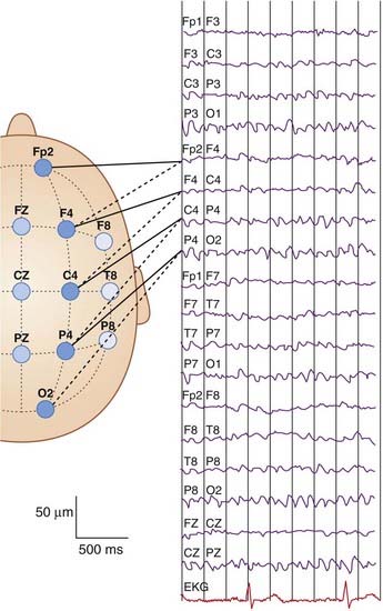
Figure 30.4 provides a complete set of normal tracings.
Types of Pattern
Normal EEG rhythms
Awake state EEG
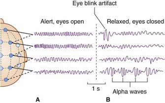
In the alert awake state (Figure 30.5A), the pattern is described as desynchronized because the waveforms are quite irregular. The background frequency is usually around 9.5 Hz. A beta frequency of more than 14 Hz may be superimposed over anterior head regions.
In a relaxed state with the eyes closed, rhythmic waveforms appear in the alpha frequency (8–14 Hz), notably over the parieto-occipital area (Figure 30.5B).
Normal sleep EEG
Glossary
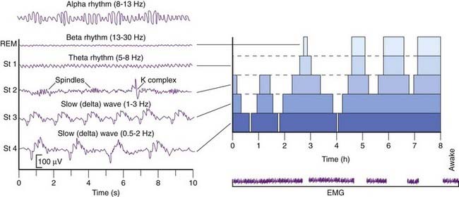
People normally pass through three to five sleep cycles per night. The sequence of events is summarized in Figure 30.5. Alpha rhythm becomes apparent (on occipital leads) during quiet rest with eyes closed.
By general agreement, sleep proper is associated with slow-wave patterns in the EEG. There is a rapid descent through stage 1, characterized by a steady theta rhythm, into stage 2, characterized by theta waves interrupted by sinusoidal waveforms called sleep spindles, and by occasional K complex spikes. Stage 3 is characterized by slow, delta waves – hence the term slow wave sleep for that stage (Figure 30.6).
As described in Chapter 27, thalamocortical projections pass through an inhibitory shell in the form of the thalamic reticular nucleus, with reciprocal connections to parent relay cells as shown in Figure 27.4
Stay updated, free articles. Join our Telegram channel

Full access? Get Clinical Tree


