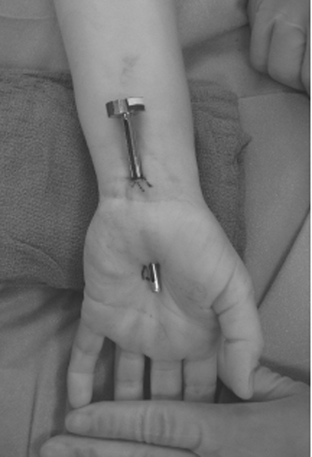
22 Endoscopic Carpal Tunnel Release A 46-year-old, right-handed female presented with a 5-year history of bilateral hand pain, greater on the right than on the left. The patient described numbness, weakness, cold sensitivity, and swelling in both hands. She described pain in the hands that often woke her at night and would be relieved by shaking her hands. Additionally, the patient stated that the pain in the right hand (8/10 in severity by a visual analog score) would radiate proximally into the forearm. Physical examination revealed atrophy of the thenar eminence on the right side and also demonstrated positive Phalen and Tinel tests bilaterally. Electromyographic studies confirmed slowing of motor conduction velocities across the carpal tunnel and sensory conduction velocities consistent with carpal tunnel syndrome. Operative options were discussed with the patient, who chose to undergo endoscopic carpal tunnel release. Biportal endoscopic release of the right median nerve was performed without complications and, in the first postoperative visit 1 week later, the patient stated that the numbness and tingling had completely resolved and the pain had decreased significantly to a level of 1 to 2/10. Furthermore, she stated that the pain and paresthesias in her right hand no longer woke her up, as they had prior to surgery. Carpal tunnel syndrome The median nerve originates from nerve roots of C6, 7, and 8, and T1. These nerve roots comprise the lateral and medial cords of the brachial plexus before forming the median nerve. The median nerve does not branch until it passes below the elbow, where in the forearm it innervates numerous wrist and digital flexors. In the hand, it supplies the “LOAF” muscles, which include the first and second lumbricales, the opponens pollicis, the abductor pollicis brevis, and the flexor pollicis brevis. The recurrent motor branch of the median nerve most commonly arises 3 cm distally to the distal wrist crease and supplies motor innervation to the abductor pollicis brevis, opponens pollicis, and superficial head of the flexor pollicis brevis. The palmar cutaneous branch of the median nerve exits the median nerve prior to its entry to the carpal tunnel and then travels alongside the median nerve superficial to the flexor retinaculum into the palm, where it divides into a medial and lateral branch supplying the skin overlying the thenar eminence. This branch typically originates ˜2 cm proximal to the upper border of the flexor retinaculum but may have a variable origin and course. The sensory supply of the median nerve is to the radial 3½ digits of the hand via the common palmar digital branches of the median nerve. The floor of the carpal tunnel is composed of the volar radiocarpal ligament and the ligamentous extensions between the carpal bones. The transverse carpal ligament, which extends over the concave surface of the carpal bones, forms the roof of the carpal tunnel. The transverse carpal ligament extends radially from the tuberosity of the scaphoid and the crest of the trapezium to its ulnar attachments, which include the hook of the hamate and the pisiform bone. Proximally, the transverse carpal ligament blends with the fibers of the antebrachial fascia at the distal wrist crease and extends ˜3 cm into the palm, near Kaplan’s cardinal line. The median nerve is located radial to the palmaris longus tendon and lies superficially beneath the transverse carpal ligament on the radial side of the tunnel. The superficialis and profundus flexor tendons lie deep within the carpal tunnel. The recurrent motor branch of the median nerve has a variety of branching patterns but the most common is the extraligamentous and recurrent type. In any branching pattern, the motor branch extends radially into the thenar muscles, which it innervates. Carpal tunnel syndrome has a well-described characteristic clinical picture. Nevertheless, many patients do not fit all of the characteristic clinical sequelae. Common presenting symptoms of this syndrome are weakness and clumsiness in the involved hand, along with numbness and paresthesias in the distribution of the median nerve. These symptoms are frequently aggravated with the use of the hand and repeated wrist flexion (e.g., brushing or combing the hair). Perhaps the most sensitive and diagnostic clinical finding involves awakening during sleep with paresthesias and numbness of the radial 3½ digits and painin the wrist and distal forearm. Greater than 90% of the patients who complain of night awakening state that shaking and rubbing the involved hand will lead to temporary resolution of the symptoms. Not uncommonly, proximal forearm, arm, and shoulder pain radiation from the wrist will be presenting symptoms. Atrophy of the median innervated muscles (e.g., thenar eminence) is a sign of an advanced median nerve entrapment neuropathy at the wrist. Although studies show greater than 50% of patients will complain of numbness and tingling along the distribution of the median nerve, ˜30% will complain of pain including all fingers and the thumb. Numerous reports conclude that motor and sensory symptoms and patient history are more important and reliable than physical examination in the diagnosis of carpal tunnel syndrome. Some reports suggest that the clinical features of carpal tunnel syndrome were more specific (66 to 87%) than sensitive (23 to 69%) for carpal tunnel syndrome. In any case, the history of repeated trauma of the hands, complaints of awakening at night with pain that is relieved by shaking of the hands, and any combination of paresthesias of the median nerve distribution, even with proximal radiation of the pain, are all compatible with carpal tunnel syndrome. Although carpal tunnel syndrome describes a classical set of symptoms and physical examination findings, it may be confused with other neurological disorders. Sensory findings along the C6 and C7 distribution may resemble a compressive radiculopathy of the C6–7 nerve roots. Patients with bilateral hand numbness and weakness (or clumsiness) should have a careful neurological examination to rule out intrinsic cervical spinal cord pathology or extrinsic compression (cervical spondylitic myelopathy). Inflammatory conditions such as arthritis in the joints, tendonitis, and fasciitis also need to be considered. Proximal forearm nerve entrapment involving the median nerve can also mimic carpal tunnel syndrome. Physical findings of muscle atrophy in the palm can certainly be mimicked by multiple neuropathies or myopathies and necessitate thorough bilateral extremities evaluation. Clinical entities that have been associated with median nerve compressive entrapment neuropathy at the wrist include pregnancy, acromegaly, diabetes, rheumatoid arthritis, hyperthyroidism, amyloidosis, gout, and alcoholism. A variety of anatomical anomalies such as ectopic muscles, vascular tumors, and ganglion cysts have been reported. The clinician must rule out cervical root compression or thoracic outlet syndrome as possible etiologies for pain in the wrist and forearm. When performing the physical examination, the examiner should be looking for symmetrical versus asymmetrical findings, involvement of other muscles, and other associated findings not following the median nerve distribution. Given that carpal tunnel syndrome is often work related in etiology, secondary gain and psychogenic issues must also be considered in all patients. A variety of studies have been performed in an attempt to predict the most useful and sensitive physical and diagnostic studies for diagnosing carpal tunnel syndrome. Although the use of nerve conduction abnormalities as a gold standard in the studies of carpal tunnel syndrome evaluation remains controversial, it is quite often used in an attempt to quantify the effectiveness of the physical examination. The Tinel sign is an examination that involves lightly tapping on the median nerve of the wrist from proximal to distal, in an attempt to reproduce tingling in the median nerve distribution. The Phalen test involves forced flexion of the wrist in an attempt to produce the paresthesias in response to this wrist position. The Durkan or carpal compression test involves the examiner pressing the carpal tunnel with his or her thumbs in an attempt to reproduce paresthesias in response to pressure within 30 seconds. Some studies have shown that the Durkan test was the most sensitive by producing positive findings in 89% of patients with electrodiagnostically proven carpal tunnel syndrome. The Phalen test and Tinel sign have sensitivity of 60% and 49%, respectively, and specificities of 80% and 55%, respectively. As stated earlier, sensory changes and weakness along the distribution of the median nerve–innervated areas are also an important part of the examination. Although the sensitivity and specificity of electrodiagnostic studies are often used as a gold standard by which other parts of history and examination are compared, there is much disagreement about how reliable these examinations are. Many studies quote their sensitivity as high as 90% or greater, whereas others argue that the sensitivities range from 49 to 84%. In any case, motor latency across the carpal tunnel of greater than 4 msec is considered diagnostic of carpal tunnel syndrome. Distal motor latencies, motor conduction velocities, and compound muscle action potential amplitudes are all variables that can be measured. Furthermore, sensory conduction velocities can be assessed as well as sensory nerve action potentials. The American Academy of Electrodiagnostic Medicine considers any variation in the measurements of greater than 2 standard deviations above or below the mean of controls to be abnormal. It is important to note when evaluating electrophysiological test results that some reports have shown a diagnosis of carpal tunnel syndrome is confirmed in only 61% of the cases by electrophysiological tests. Many of these patients with negative studies still receive significant relief from surgical intervention. Furthermore, some patients with severe carpal tunnel syndrome proven by diagnostic studies will achieve symptomatic relief, particularly to pain, with operative intervention, whereas electrodiagnostic follow-ups may show no change in their findings. The full gamut of options for open carpal tunnel release have been presented in Chapter 2. After the diagnosis of carpal tunnel syndrome is made, the first line of treatment consists of conservative nonsurgical therapy. Although not commonly possible or feasible, repetitive stressful motion of the hands should be curtailed. Splinting the affected wrist in neutral position typically relieves the symptoms in up to 80% of the patients. However, in the majority of these patients, the symptoms return once the splints are removed. Some physicians treat patients with steroid injections, which have also been found to have limited success. When the etiology of carpal tunnel syndrome is temporary and self-correcting (i.e., pregnancy) conservative therapy is the treatment of choice. If the patient fails an adequate trial of conservative therapy, and electrodiagnostic studies demonstrate progressive neuronal/axonal dysfunction, then the patient is deemed a candidate for surgical decompression, which may be done via an open or endoscopic approach. Treatment of carpal tunnel syndrome with endoscopic techniques was first begun in 1989, and six types of procedures have been developed since. Three types use a single incision (uniportal) and three types use two small incisions (biportal). Described herein is the author’s preferred method, which is the biportal technique as described by Brown. Although the procedure can be performed under local anesthesia or using a Bier block, our preferred method of choice is to induce the patient using anesthesia consisting of an intravenous infusion of propofol along with a laryngeal mask airway. Following application of the tourniquet, a 1 cm incision is made 1 to 2 cm proximal to the distal wrist crease immediately ulnar to the palmaris longus tendon. The antebrachial fascia is then exposed and bluntly divided. A synovial elevator is advanced distally under the antebrachial fascia and effortlessly into the carpal tunnel. The synovium is removed from the undersurface of the transverse carpal ligament. An obturator with a slotted cannula is inserted into the carpal tunnel and exited distally ˜4 cm distal to the distal wrist crease along the third web space (Fig. 22–1). The obturator is removed and a 30 degree rigid endoscope is inserted distally and used to visualize the undersurface of the transverse carpal ligament through the slotted end of the cannula (Fig. 22–2). A hook blade is then inserted proximally and advanced distally to the distal end of the transverse carpal ligament. Under direct visualization, the ligament is divided in its entirety from distal to proximal on the ulnar side of the ligament next to the hook of the hamate (Fig. 22–3). Depending on the thickness of the ligament, several passes may be required to fully resect the ligament. No attempt is made to visualize the median nerve. Following tourniquet deflation, hemostasis is obtained, the incisions are closed with three simple nylon sutures, and a volar splint is applied. The sutures and the splint are removed within 5 to 7 days. The patients typically return to work 2 weeks following endoscopic release.
 Case Presentation
 Diagnosis
 Anatomy
 Characteristic Clinical Presentation
 Differential Diagnosis
 Diagnostic Tests
 Management Options
 Endoscopic Surgical Release

Stay updated, free articles. Join our Telegram channel

Full access? Get Clinical Tree


