Enhancing Suprasellar Mass
Anne G. Osborn, MD, FACR
DIFFERENTIAL DIAGNOSIS
Common
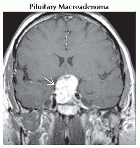
Pituitary Macroadenoma
Meningioma
Saccular Aneurysm
Craniopharyngioma
Pilocytic Astrocytoma
Less Common
Diffuse Astrocytoma, Low Grade
Neurosarcoid
Langerhans Cell Histiocytosis
Germinoma
Lymphocytic Hypophysitis
Rare but Important
Metastasis
Lymphoma
Leukemia
ESSENTIAL INFORMATION
Key Differential Diagnosis Issues
Effect of age on differential diagnosis important
Common lesions (“big five”) account for > 75% of all suprasellar masses
Most other lesions < 1-2% each
Differential diagnosis narrows if mass confined to infundibulum
Helpful Clues for Common Diagnoses
Macroadenoma vs. Meningioma
Macroadenoma: Gland can’t be identified separate from mass
Meningioma: Mass distinct from gland (hypointense diaphragma sellae separates mass above from pituitary gland below)
Saccular Aneurysm
Coronal plane helpful in distinguishing aneurysm from normal pituitary
Look for phase artifact, “flow void” on MR
Craniopharyngioma
90% Ca++, 90% cystic, 90% enhance
Papillary variant (more common in adults) may be solid, noncalcified, enhances strongly
Pilocytic Astrocytoma
More common in children
Expands hypothalamus, optic chiasm, may extend into optic nerves/tracts
Variable enhancement
Pilomyxoid variant
Infant > child
Aggressive behavior
Large, bulky tumor with lateral extension to temporal lobe common
Hemorrhage in 20-25%
Helpful Clues for Less Common Diagnoses
Histiocytosis, germinoma in children/young adults
Neurosarcoid in older patients
Helpful Clues for Rare Diagnoses
Solitary metastasis to gland/stalk rare
Lymphoma, leukemia usually with systemic disease
Image Gallery
 Coronal T1 C+ MR shows inhomogeneously enhancing intra- and suprasellar mass
 . Pituitary gland can’t be found separate from and indeed IS the mass. . Pituitary gland can’t be found separate from and indeed IS the mass.Stay updated, free articles. Join our Telegram channel
Full access? Get Clinical Tree
 Get Clinical Tree app for offline access

|