Enlarged Pituitary Gland
Anne G. Osborn, MD, FACR
DIFFERENTIAL DIAGNOSIS
Common
Pituitary Hyperplasia
Pituitary Microadenoma
Pituitary Macroadenoma
Less Common
Neurosarcoid
Langerhans Cell Histiocytosis
Lymphocytic Hypophysitis
Pituitary Macroadenoma (Mimic)
Rare but Important
Intracranial Hypotension
Meningioma
Metastases to Gland/Stalk
Dural A-V Fistula
Pituicytoma
Pseudotumor, Intracranial
Lymphoma, Primary CNS
Leukemia
ESSENTIAL INFORMATION
Key Differential Diagnosis Issues
Not all “enlarged pituitary glands” are abnormal!
Size/height varies with gender, age
Children = 6 mm
Males, postmenopausal females = 8 mm
Young menstruating females = 10 mm (can bulge upwards)
Pregnant, postpartum lactating females = 12 mm
Enhances strongly, uniformly
15-20% have incidental cyst or nonfunctioning microadenoma (pituitary “incidentaloma”)
Variants/mimics of “enlarged pituitary”
“Pseudoenlargement” secondary to unusually shallow bony sella
Medially positioned cavernous internal carotid arteries (“kissing carotids”) may make gland appear enlarged
Helpful Clues for Common Diagnoses
Pituitary Hyperplasia
Can be normal (young menstruating females)
Enlarged gland ± upward bulging
May be related to end-organ failure or neuroendocrine tumors
Pituitary Microadenoma
May enlarge gland
Best identified with dynamic, contrast-enhanced MR
Pituitary Macroadenoma
Pituitary gland can’t be distinguished from mass
Enhances strongly, often heterogeneously
Other Essential Information
Venous congestion (intracranial hypotension, dAVF) can enlarge gland
Beware: Child or young adolescent male with “pituitary adenoma” most likely has pituitary hyperplasia, not neoplasm!
Evaluate for end-organ failure (e.g., hypothyroidism)
Image Gallery
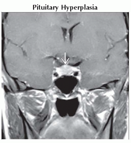 Coronal T1 C+ MR shows a physiologically enlarged pituitary gland
 in this 28 year old lactating woman. The gland measures nearly 12 mm in height. Follow-up scan 1 year later was normal. in this 28 year old lactating woman. The gland measures nearly 12 mm in height. Follow-up scan 1 year later was normal.Related posts:Stay updated, free articles. Join our Telegram channel
Full access? Get Clinical Tree
 Get Clinical Tree app for offline access

|