SECTION I BASIC PRINCIPLES
CHAPTER 1
Fundamentals of the Nervous System
More than any other organ, the nervous system makes human beings special. The human central nervous system (CNS), smaller and weighing less than most desktop computers, is the most complex and elegant computing device that exists. It receives and interprets an immense array of sensory information, controls a variety of simple and complex motor behaviors, and engages in deductive and inductive logic. The brain can make complex decisions, think creatively, and feel emotions. It can generalize and possesses an elegant ability to recognize that cannot be reproduced by even advanced computers. The human nervous system, for example, can immediately identify a familiar face regardless of the angle at which it is presented. It can carry out many of these demanding tasks in a nearly simultaneous manner.
Given the complexity of the nervous system and the richness of its actions, one might ask whether it can ever be understood. Indeed, neuroscience has begun to provide an understanding, in elegant detail, of the organization and physiology of the nervous system and the alterations in nervous system function that occur in various diseases. This understanding is firmly based on an appreciation of the structure of the nervous system and the interrelation between structure and function.
The complexity of the nervous system’s actions is reflected by a rich and complex structure—in a sense, the nervous system can be viewed as a complex and dynamic network of interlinked computers. Nevertheless, the anatomy of the nervous system can be readily understood. Since different parts of the brain and spinal cord subserve different functions, the astute clinician can often make relatively accurate predictions about the site(s) of dysfunction on the basis of the clinical history and careful neurological examination. An understanding of neuroanatomy is immediately relevant to both basic neuroscience and clinical medicine. Clinical neuroanatomy (i.e., the structure of the nervous system, considered in the context of disorders of the nervous system) can teach us important lessons about the structure and organization of the normal nervous system, and is essential for an understanding of disorders of the nervous system.
GENERAL PLAN OF THE NERVOUS SYSTEM
Main Divisions
A. Anatomy
Anatomically, the human nervous system is a complex of two subdivisions.
1. CNS—The CNS, comprising the brain and spinal cord, is enclosed in bone and wrapped in protective coverings (meninges) and fluid-filled spaces.
2. Peripheral nervous system (PNS)—The PNS is formed by the cranial and spinal nerves (Fig 1–1).
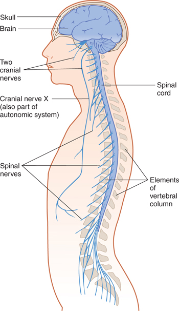
FIGURE 1–1 The structure of the central nervous system and the peripheral nervous system, showing the relationship between the central nervous system and its bony coverings.
B. Physiology
Functionally, the nervous system is divided into two systems.
1. Somatic nervous system—This innervates the structures of the body wall (muscles, skin, and mucous membranes).
2. Autonomic (visceral) nervous system (ANS)—The ANS contains portions of the central and peripheral systems. It controls the activities of the smooth muscles and glands of the internal organs (viscera) and the blood vessels and returns sensory information to the brain.
Structural Units and Overall Organization
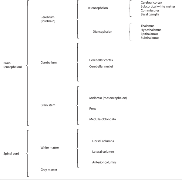
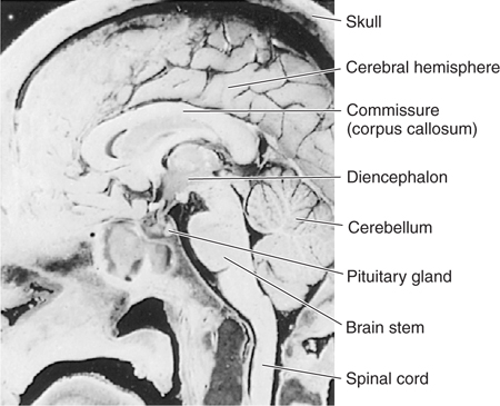
The central portion of the nervous system consists of the brain and the elongated spinal cord (Fig 1–2 and Table 1–1). The brain has a tiered structure and, from a gross point of view, can be subdivided into the cerebrum, the brain stem, and the cerebellum.
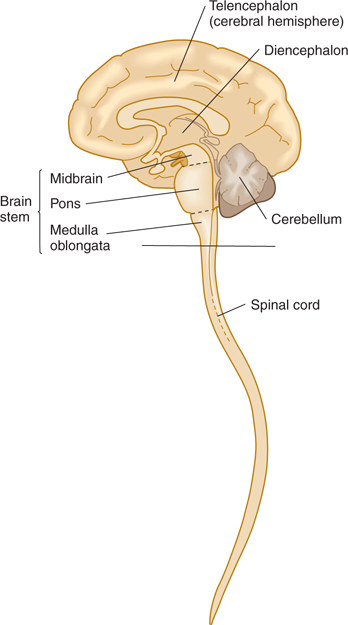
FIGURE 1–2 The two major divisions of the central nervous system, the brain and the spinal cord, as seen in the midsagittal plane.
TABLE 1–1 Major Divisions of the Central Nervous System.
The most rostral part of the nervous system (cerebrum, or forebrain) is the most phylogenetically advanced and is responsible for the most complex functions (eg, cognition). More caudally, the brain stem, medulla, and spinal cord serve less advanced, but essential, functions.
The cerebrum (forebrain) consists of the telencephalon and the diencephalon; the telencephalon includes the cerebral cortex (the most highly evolved part of the brain, sometimes called “gray matter”), subcortical white matter, and the basal ganglia, which are gray masses deep within the cerebral hemispheres. The white matter carries that name because, in a freshly sectioned brain, it has a glistening appearance as a result of its high lipid-rich myelin content; the white matter consists of myelinated fibers and does not contain neuronal cell bodies or synapses (Fig 1–3). The major subdivisions of the diencephalon are the thalamus and hypothalamus. The brain stem consists of the midbrain (mesencephalon), pons, and medulla oblongata. The cerebellum includes the vermis and two lateral lobes. The brain, which is hollow, contains a system of spaces called ventricles; the spinal cord has a narrow central canal that is largely obliterated in adulthood. These spaces are filled with cerebrospinal fluid (CSF) (Figs 1–4 and 1–5; see also Chapter 11).
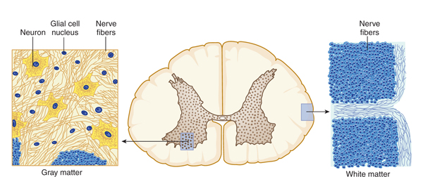
FIGURE 1–3 Cross section through the spinal cord, showing gray matter (which contains neuronal and glial cell bodies, axons, dendrites, and synapses) and white matter (which contains myelinated axons and associated glial cells). (Reproduced, with permission, from Junqueira LC, Carneiro J, Kelley RO: Basic Histology: Text & Atlas, 11th ed. McGraw-Hill, 2005.)
Stay updated, free articles. Join our Telegram channel

Full access? Get Clinical Tree


