Fundoscopy
Examining the fundus with an ophthalmoscope is often thought to be particularly difficult. This need not be the case with an understanding of how to use an ophthalmoscope, a clear idea of what is normal and what are normal variants, the appearances of common and important abnormalities and, of course, practice.
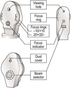
Setting up the ophthalmoscope
Set focus ring to 0 (Fig. 1). Remove dust cover. Select beam to plain. Some people remove their own glasses; if so then move the focus ring to correct. Make sure the light works.


Sit the patient comfortably and ask him or her to look at a particular point in the distance. Draw curtains, turn down lights. With your eye in the same plane as the patient’s eye and the fixation point, and at 15° from the line between eye and point of fixation (so you are aiming at the centre of the occiput), look at the eye from about 30 cm (Fig. 2). Look at the red reflex. Any lens or vitreous opacity will be seen in silhouette. Gradually move in towards the eye. The disc should come into view. The ophthalmoscope may need focusing. If the disc is not seen, find a vessel and track it from branches to trunk.
Stay updated, free articles. Join our Telegram channel

Full access? Get Clinical Tree


