Fig. 1.1
Title page of Stephanus ’ De dissectione partium corporis humani (1545) (Courtesy History & Special Collections for the Sciences, Louise M. Darling Biomedical Library, University of California, Los Angeles)
1.1.2 Nomenclature and Terminology
The term syringomyelia, describing a tube-like cavity within the spinal cord, was first applied in 1827 by Ollivier d’Angers (Fig. 1.2). He thought that the central canal of the spinal cord was not a normal finding. In 1865, Schüppel described a case of hydromyelia as an upward expansion of the central canal, which then ended in a cavity. By 1890, it was recognised that the central canal is always present (Bruhl 1890). Simon (1875) preferred the term syringomyelia as being more general than hydromyelia, which he defined as hydropic widening of the central canal. For many years the distinction between syringomyelia and hydromyelia became blurred, leading to the invention of terms such as “hydrosyringomyelia” or “syringohydromyelia”. Modern imaging techniques have drawn attention to persistence of the central canal as a non-pathological finding, which is best termed hydromyelia. Some have suggested that persistence of the central canal in patients who sustain spinal cord injury or in individuals who have tonsillar ectopia, may explain the development of syringomyelia in these individuals, while others with cord injuries or tonsillar ectopia do not develop syringomyelia (Milhorat et al. 1994).
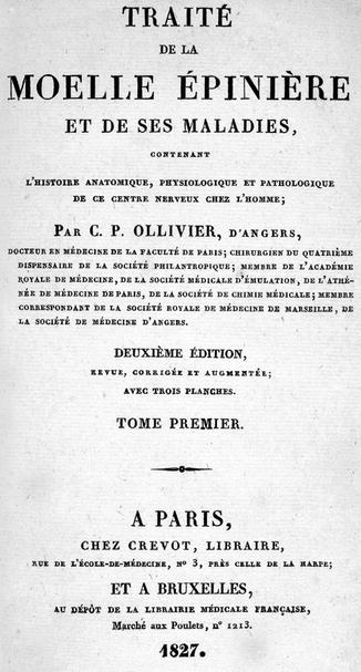
Fig. 1.2
Title page of Ollivier’s Traité de la Moelle Épiniere (1827) (Courtesy History & Special Collections for the Sciences, Louise M. Darling Biomedical Library, University of California, Los Angeles)
Schϋppel also cited the case of Brunner’s in which there was communication with the fourth ventricle. We now appreciate that communication between spinal cord cavities and the fourth ventricle is present in only a small number of cases (West and Williams 1980). The once-presumed communication between a syrinx cavity and the fourth ventricle in patients with cerebellar tonsillar ectopia also led to the now-abandoned distinction between “communicating” and “non-communicating” forms of syringomyelia (Barnett et al. 1973b). On the other hand, isolated spinal cord cavities rarely, but occasionally, do communicate with the subarachnoid space (Milhorat et al. 1995).
1.1.3 First Descriptions of Tonsillar Ectopia
In 1881 Theodor Langhans described tonsillar ectopia and suggested that, by obstructing flow at the foramen magnum, it might result in syringomyelia. In 1883 Cleland described nine infants with spina bifida who had various cerebral anomalies, including hydrocephalus and anencephalus. Elongation of the cerebellar tonsils is evident in his figure 6 (specimen 1). In 1891 Chiari provided his first description of hindbrain abnormalities in association with hydrocephalus. His 1896 publication was more detailed and focused specifically on changes in the cerebellum, pons and medulla. He described four abnormalities, differing in degree of cerebellar abnormality (Table 1.1). In 1894 Arnold added a case of an infant with tonsillar descent below the foramen magnum. In 1943 Lichtenstein, a pathologist, also postulated that there was a relationship between tonsillar descent and syringomyelia. We now recognise that there are two general categories of syringomyelia: (a) syringomyelia associated with tonsillar descent (Chiari malformation) and (b) primary spinal syringomyelia, in which the pathology is entirely confined to the spinal cord and its meninges (Williams 1991).
Table 1.1
The four varieties of Chiari malformations
Type I: Downward displacement of the cerebellar tonsils and the medial portion of the inferior cerebellar lobes |
Type II: Downward displacement of the tonsils, vermis and at least a part of a lengthened fourth ventricle |
Type III: Downward displacement of [nearly] the entire cerebellum, out of the cranial cavity, into the cervical area |
Type IV: Hypoplasia of the region of the cerebellum without displacement of this structure into the spinal canal |
1.1.4 Other Forms of Syringomyelia
Strümpel (1880) may have been the first to identify a case of syringomyelia after trauma. This condition was also recognised by Schlesinger (1895), but the major description was provided by Barnett (Barnett 1973a; Barnett et al. 1973b). Barnett (1973b) also noted that delayed syringomyelia might occur after both severe and less severe spinal injury. Earlier discussions and treatises on syringomyelia also tended to include spinal cord tumour cysts under the broad category of syringomyelia (Barnett and Rewcastle 1973). While spinal cord tumours may be associated with true syringomyelia cavities, notably when the tumour obstructs or narrows the spinal subarachnoid space, syrinx cavities need to be distinguished from tumour cysts, which are often high in protein and represent a different entity, from both physiological and treatment perspectives.
1.2 Elucidation of Clinical Features
Aspects of the motor and sensory deficits caused by syringomyelia had been defined by the end of the nineteenth century, permitting the diagnosis of syringomyelia in a living patient (Kahler and Pick 1879; Schultze 1882). In 1869 Charcot described dissociated anaesthesia with absent upper extremity reflexes and atrophy in one or both upper limbs, particularly the hands. He also described the severe joint deformities, especially the shoulder joints, which characteristically may occur in areas of absent pain sensation also involving the upper trunk (Charcot 1868). Duchenne first called attention to muscular atrophy in association with sensory abnormalities in 1853 and differentiated this condition from the muscular dystrophy that bears his name. Gowers, in his 1886 textbook, provided a more detailed description of the various clinical manifestations of syringomyelia. Milhorat et al. (1999) compiled a comprehensive list of symptoms linked with syringomyelia associated with tonsillar ectopia.
The motor and sensory deficits encountered in patients with primary spinal syringomyelia also relate to the level and degree of spinal cord injury. Foster and Hudgson (1973) described the sometimes-sudden ascent of the sensory level in patients with post-traumatic syringomyelia, not infrequently precipitated by spells of coughing or straining. They also called attention to the fact that it may be difficult to distinguish deficits due to the cord injury from those due to syrinx formation.
1.3 Theories of Pathogenesis
Many theories have surfaced over the course of the years, relating to the origin of syringomyelia cavities. These have implicated inflammation, including specific diseases such as syphilis and arachnoiditis, possibly leading to glial proliferation (Hallopeau 1870), with subsequent degenerative changes resulting in cavity formation (Schultze 1882). A variety of other mechanisms have since been proposed, including congenital abnormalities, neoplasia, ischaemia and processes leading to oedema of the cord (Klekamp and Samii 2002).
The observations of Cleland (1883) and of Chiari (1891, 1896) suggested to them that syrinx cavities fill from the fourth ventricle. Based on this concept, Gardner and Angel (1958) developed the theory of a water hammer effect as the filling mechanism of syrinx cavities. Ellertson and Greitz’s (1939) studies tended to strengthen this concept of syrinx communication with the fourth ventricle, but their fluorescein dye experiments also raised the possibility of transparenchymal fluid migration from the subarachnoid space. Ball and Dayan (1972) suggested that fluid enters the syrinx cavity via the Virchow-Robin spaces during Valsalva manoeuvres, propelled by epidural venous distension. The cerebellar tonsils were postulated to prevent upward propagation of the CSF pulse wave. Brierley (1950) injected a suspension of India ink into the subarachnoid space of rabbits and noted some of this material in the perivascular spaces of the cord. Studies by Rennels et al. (1985) with horseradish peroxidase established fluid flow into the tissues of the neuraxis by a “paravascular” pathway. Stoodley et al. (1996) was able to demonstrate that fluid migrates into the cord along the Virchow-Robin spaces, evidently propelled by the pulsation of arterioles in these spaces. Milhorat et al. (1994) suggested that persistence of the central canal of the spinal cord might play a role in the likelihood of syrinx formation. A currently widely accepted theory proposed by Oldfield is that systolic pressure waves cause the impacted tonsils to act as pistons exerting pressure on the relatively closed spinal subarachnoid space, thereby driving fluid into the spinal cord (Oldfield et al. 1994).
Expansion of the cystic cavity, once formed, also raised questions. Both Barnett et al. (1973a) and Williams (1970, 1991) considered venous expansion in the area below the injury as a significant factor.
Spinal haemorrhage and necrosis were, at one time, believed to be the basis of post-traumatic syringomyelia (Barnett et al. 1973a). Diffusion of fluid from blood vessels was also suggested. Barnett (1973b) also observed that syrinx formation might occur following relatively minor spinal injuries not necessarily associated with immediate neurological impairment, thereby raising doubts about these theories. Partial obstruction of the subarachnoid space is now considered to be the underlying pathophysiology of many forms of primary spinal syringomyelia (Batzdorf 1991). Arachnoid scarring, sometimes referred to as arachnoiditis in the older literature, is believed to act in a manner analogous to how the cerebellar tonsils behave in Chiari-related syringomyelia, preventing unimpaired transmission of the CSF pulse wave along the spinal subarachnoid space. Alterations in the cord parenchyma due to injury may facilitate inflow of CSF into the cord. Focal membranes, such as are seen in arachnoid cysts , are believed to result in syringomyelia by a similar mechanism (Holly and Batzdorf 2006).
1.4 Development of Imaging Methods
The development of methods of imaging a syrinx cavity, even though it was only visualised indirectly, represented a major advance over localisation by neurological examination alone. Thus, a radiographic finding of bony spinal canal expansion was a useful aid to the diagnosis of suspected syringomyelia in a patient with appropriate clinical signs or symptoms, even before oil contrast myelography allowed one to visualise the expanded spinal cord (Boijsen 1954). Imaging the syringomyelia cord with oxygen was described in 1949 (Marks and Livingston 1949). Variability in cord diameter on air myelography , in relation to patient position, was reported in 1966 (Westberg 1966). Pantopaque myelography, demonstrating a distended cervical spinal cord, followed by air myelography with the patient in upright position, permitted demonstration of collapse of a distended cord in relation to changes in body position (Conway 1967). Real advances were, however, possible only after the introduction of water-soluble contrast material. Experience with computerised tomography (CT) following instillation of such media was reported in 1980 (Aubin et al. 1981). This included the significant observation that contrast could be imaged within the syrinx cavity on delayed CT scans. Magnetic resonance (MR) imaging, in addition to showing the cord cysts in great detail and allowing the demonstration of tumours, had the great advantage of not being invasive. By comparison, myelography required needle puncture of the theca, potentially changing the fluid dynamics within the spinal CSF channels. A very early MR image was published in 1983 (Batnitzky et al. 1983), and many refinements in technique over the ensuing years have yielded the exquisitely detailed studies currently available. MR technology has also provided insights into the physiology of syringomyelia (Enzmann and Pelc 1989). Constructive interference in steady-state MR images (CISS ) is an example of recent refinements, with superior visualisation of subarachnoid webs that may indicate potential benefits from surgery (Korogi et al. 2000; Roser et al. 2010). Future advances in technology will undoubtedly add additional information.
A further account of the history of imaging of syringomyelia is provided in Chap. 21 of this publication.
1.5 Treatment
The earliest attempts at treatment of syringomyelia were directed at relieving the fluid collection within the spinal cord, beginning in 1892, with open cyst aspiration (Abbe and Coley 1892), followed by myelotomy in 1921 (Elsberg 1921) and 1926 (Poussepp 1926) and insertion of a drain into the cystic cavity in 1936 (Frazier and Rowe 1936). Percutaneous cyst aspiration was described in 1966 (Westberg 1966). Tantalum drains and plastic tubing were placed in 1949 (Kirgis and Echols 1949). The use of Pantopaque (Myodil ) by injection to obliterate the cyst was proposed in 1981 (Schlesinger et al. 1981). Penfield and Coburn’s patient of 1938, although often cited, evidently had a Chiari malformation without syringomyelia.
Noting that in the few cases in which fluid had been surgically evacuated, there was no significant effect on the condition, and also on the basis that syrinx cavities seemed to be caused by glial proliferation, radiation therapy was proposed in 1905 (Raymond 1905). As late as 1955, it was stated in Brain’s textbook that radiation therapy was the generally accepted form of treatment of syringomyelia.
Recognising the coexistence of hydrocephalus and syringomyelia, Bernini and Krayenbühl (1969) suggested ventricular shunting of such patients. Although of interest from a theoretical point of view, the results were disappointing. More local diversion of CSF in the spinal subarachnoid space has shown somewhat better results (Vengsarkar et al. 1991; Lam et al. 2008). Technical improvements in syrinx drainage followed, notably syrinx-to-peritoneal shunting (Edgar 1976), subarachnoid shunting (Tator et al. 1982; Isu et al. 1990; Iwasaki et al. 1999) and syrinx-to-pleural cavity shunting (Williams and Page 1987). Gardner et al.’s (1977) novel concept of syrinx drainage by performing a “terminal ventriculostomy ” was unsuccessful in many patients, in large part because it did not address the filling mechanism of syrinx cavities (Williams and Fahy 1983).
Related posts:


Stay updated, free articles. Join our Telegram channel

Full access? Get Clinical Tree


