Fig. 45.1
MRI and CT findings in a 10-year-old-girl with a clival chordoma. Sagittal T1-weighted MR image shows a clival mass that has a hyperintense focus which represents highly proteinaceous material or blood products (a). Axial T1-weighted MR image shows an isointense clivus mass with hyperintense focus (b). Axial CT scan demonstrates lytic bone destructions with intratumoral bone or calcifications (c). Contrast-enhanced axial CT scan shows irregular tiny enhancement (d)

Fig. 45.2
MRI and CT findings in an 11-year-old-boy with a clival chordoma. Sagittal T2-weighted image represents a clivus mass with irregular tiny septa, extending to the upper cervical spine (a). There is extensive brainstem compression. Axial postcontrast CT scan demonstrates a clivus mass with a little enhancement (b)


Fig. 45.3
CT, MRI, and angiographic findings in an 18-year-old woman with a skull base chordoma. Axial fat saturation T2-weighted image (a) shows a parasagittal mass extending to the sphenoid sinus. The mass is hyperintense and contains hypointense focus compatible with bone remnant or calcifications. The mass displaces and compresses the left internal carotid artery anteromedially (a, k). Axial unenhanced T1-weighted image demonstrates the border of the tumor (b). There is marked enhancement on contrast-enhanced fat-saturated T1-weighted image which also delineates the border of the tumor (d). Axial GRE T2* image depicts the calcifications better than the other sequences (c). Axial diffusion-weighted image and corresponding ADC map reveal an increase in diffusion (e, f). The soft and bone tissue algorithm of axial CT scan demonstrate the border and contents of the tumor (h, i). Axial collapsed MIP image of 3D TOF MR angiography demonstrates the displacement in internal carotid artery (g). Left internal carotid arteriogram confirms the vascular displacement (j). There is no staining of the tumor in the arterial phase

Fig. 45.4
MR findings of a 5-year-old-boy with clivus chordoma. Sagittal T2-weighted image demonstrates an intracranial chordoma with slight irregular hyperintensity due to tiny hypointense septa (a). The huge mass extends to the upper cervical spine and compresses the brainstem posteriorly (a, b). Axial contrast-enhanced T1-weighted image shows a large clival mass with variable enhancement (honeycomb enhancement pattern) (c)
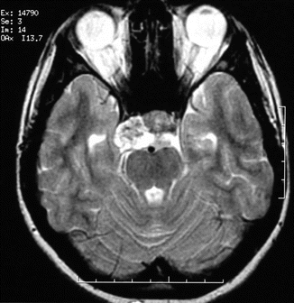
Fig. 45.5
Axial T2-weighted image of a 17-year-old girl with clivus chordoma shows a heterogeneous mass in the upper part of the clivus extending to the right cavernous sinus
45.1 Computed Tomography
CT scanning is essential, highly sensitive, and accurate for evaluating bony integrity, bone destruction, and calcifications or bone fragments within the lesion [3, 6]. CT should be reviewed in both soft and bone algorithm with multiplanar reconstruction (Fig. 45.3h, i). The classic appearance of the clivus chordomas at CT is that of a centrally located, well-circumscribed expansile soft-tissue mass with extensive lytic bone destruction [12] (Fig. 45.2b). The margin between the tumor and adjacent bone is not sclerotic. CT easily defines the exact nature and extent of the bone destruction caused by the tumor. CT is valuable in evaluating the integrity of cranio-vertebral junction and reliably demonstrates the lysis of the skull base foramina. The bulk of the tumor is usually hyperattenuating relative to the neighboring neural axis. Intratumoral calcifications appear irregular (Figs. 45.1c and 45.3h). Moderate to marked enhancement following administration of iodinated contrast material is seen in almost every case (Fig. 45.1d). Solitary or multiple low-attenuation areas are sometimes seen within the soft-tissue mass representing gelatinous material seen at gross examination (Fig. 45.2b). The bone changes and the extent of the resection after operation are precisely interpreted at CT [1].
45.2 MRI
Clivus chordomas have intermediate to low signal intensity and is easily recognized within the high signal intensity of the fat of the bone marrow of clivus on T1-weighted MR images (Figs. 45.1a, b, 45.3b and 45.4b). Small foci of hyperintensity can be visualized within the tumor on T1-weighted images which may represent a protein-rich content or intratumoral hemorrhage [1, 13] (Fig. 45.4b). Typically, intracranial chordoma has high signal intensity on T2-weighted images due to the high fluid content of cellular components (Figs. 45.2a and 45.4a). The intratumoral areas of calcification, hemorrhage, and a highly proteinaceous mucus pool usually demonstrate heterogeneous hypointensity at T2-weigted images (Fig. 45.3a, c). Low-signal-intensity irregular septations that separate high-signal-intensity lobules are commonly seen (Figs. 45.2a and 45.4a). MRI provides accurate evaluation of the involvement of the adjacent soft tissues, including vasculature, cranial nerve involvement, and changes in the brainstem related to tumor itself [1, 2, 6] (Figs. 45.3a and 45.4a). Fat saturation with T2-weighted images can be used to increase in delineation of the border of the tumor (Fig. 45.3a). Gradient echo T2* or susceptibility-weighted images (SWI) may show intratumoral calcification or hemorrhage better than conventional T1- and T2-weighted images (Fig. 45.3c). Furthermore, SWI can make a differentiation between calcification and hemorrhage with phase reconstruction algorithm.
Related posts:


Stay updated, free articles. Join our Telegram channel

Full access? Get Clinical Tree


