10 Innervation of muscles and joints
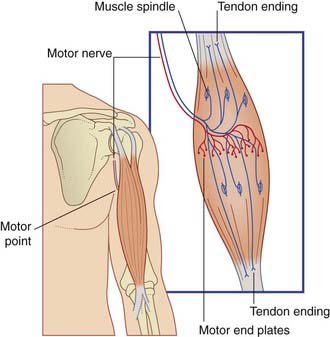
In gross anatomy, the nerves to skeletal muscles are branches of mixed peripheral nerves. The branches enter the muscles about one-third of the way along their length, at motor points (Figure 10.1). Motor points have been identified for all major muscle groups for the purpose of functional electrical stimulation by physical therapists, in order to increase muscle power.
Motor Innervation of Skeletal Muscle
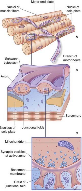
The nerve of supply branches within the muscle belly, forming a plexus from which groups of axons emerge to supply the muscle fibers (Box 10.1 and Figure 10.1). The axons supply single motor end plates placed about halfway along the muscle fibers (Figure 10.2A).
There are three different types of skeletal muscle fiber.
Motor end plates
At the myoneural junction, the axon divides into a handful of branchlets that groove the surface of the muscle fiber (Figure 10.2B). The underlying sarcolemma is thrown into junctional folds. The basement membrane of the muscle fiber traverses the synaptic cleft and lines the folds. The underlying sarcoplasm shows an accumulation of nuclei, mitochondria, and ribosomes known as a sole plate.
Each axonal branchlet forms an elongated terminal bouton containing thousands of synaptic vesicles loaded with acetylcholine (ACh). Synaptic transmission takes place at active zones facing the crests of the junctional folds (Figure 10.2C). Vesicular ACh is extruded at great speed by exocytosis into the synaptic cleft. The ACh diffuses through the basement membrane to bind with ACh receptors in the sarcolemma.
Also in terminal boutons are some dense-cored vesicles containing one or more peptides (Figure 12.2C). Best known is calcitonin gene-related peptide, a potent vasodilator.
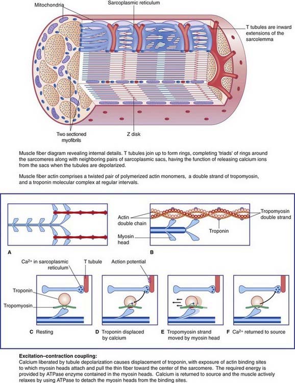
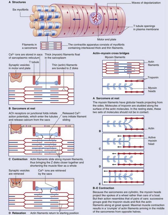
Details of the muscle fiber contraction process are in Box 10.2.