Chapter 17 Intradiscal Therapies
Intradiscal decompression for chronic intractable discogenic pain can be achieved by means of various methods, such as intradiscal electrothermal therapy (IDET), nucleoplasty, and percutaneous discectomy. Tables 17.1 and 17.2 summarize these three techniques.
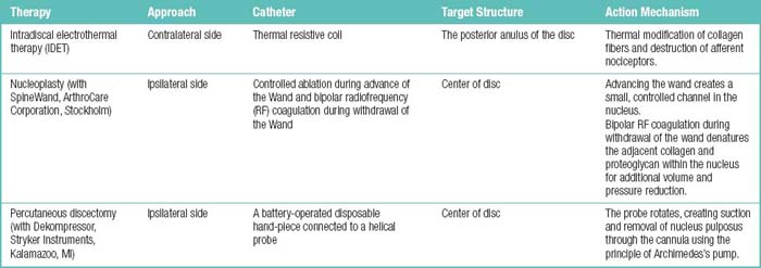
Table 17.2 The Evidence for Efficacy Intradiscal Electrothermal Therapy (IDET) and Nucleoplasty in Management of Chronic Discogenic Low Back Pain
| Therapy | Evidence of Short-Term Relief | Evidence of Long-Term Relief |
|---|---|---|
| IDET | Strong | Moderate |
| Nucleoplasty | Limited | Limited |
Data from Boswell MV, Shah RV, Everett CR, et al. Interventional techniques in the management of chronic spinal pain: Evidence-based practice guidelines. Pain Physician 2005;8:1-47.
Intradiscal electrothermal therapy
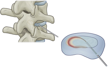
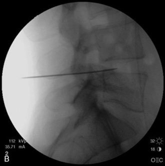
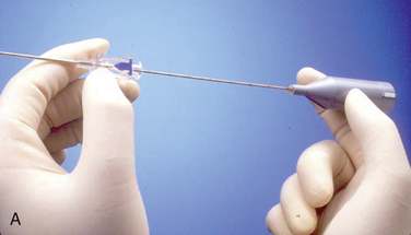
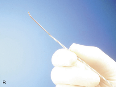
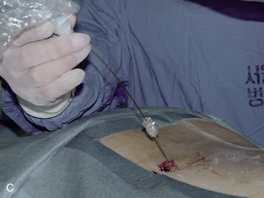
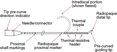
In IDET, the collagen fibers of the disc are modified and the nociceptors causing axial back pain are destroyed by thermal energy delivered through the percutaneous threading of a flexible catheter, composed of a thermal resistive coil, into the disc from the side contralateral to the lesion under fluoroscopic guidance (Fig. 17-1).
Mechanisms of Action
Thermal modification of collagen fibers acts as follows:

 With disc temperatures reaching 65° C, collagen may contract as much as 35% from its original size (Fig. 17-2).

Indications and Contraindications
Ideal candidates for IDET have the following features:


Indications
The indications for IDET are as follows:


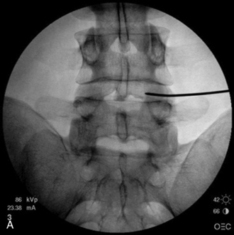
 At the target level, the discogram and subsequent computed tomography scanning demonstrate spread of the contrast agent to the outer anulus or beyond the confines of the disc (Fig. 17-3)

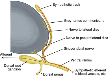


Related Anatomy and Pathophysiology
Figure 17-3 illustrates innervations of an intervertebral disc related to IDET.
Procedure


Stay updated, free articles. Join our Telegram channel

Full access? Get Clinical Tree


