♦ Preoperative
Operative Planning
- Magnetic resonance imaging (MRI): enhancement, syrinx, edema
- Define rostral/caudal extent of tumor
- Computed tomography: spinal dysraphism
- Clinical: history of lipomyelomeningocele repair; look for hydrocephalus
- If lumbar, bladder/bowel function requires formal assessment
Equipment
- Basic spine tray
- Drill
- 1-, 2-, and 3-mm Kerrison rongeurs
- Operating microscope
- Somatosensory evoked potentials and motor evoked potentials; electromyography in cauda equina lipoma
- Ultrasonic aspirator
♦ Intraoperative
Approach
- Dictated by level of involvement: cervical, thoracic, lumbar
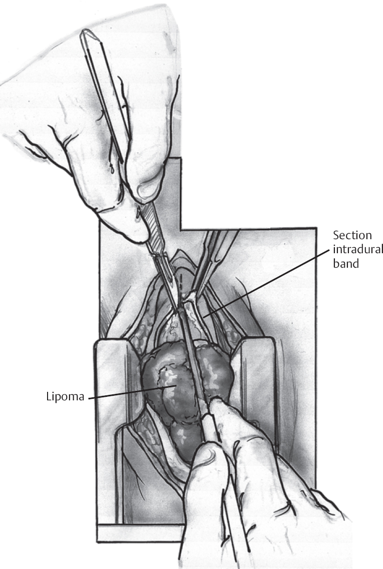
Tumor Resection (Fig. 134.1)
- Dexamethasone 10 mg or 20 mg intravenous at start of case
- Standard laminectomy with patient prone
- Consider laminoplasty if possible
- Dura opened midline and tented to muscle laterally
- Identify dorsal midline by visualizing exiting nerve roots bilaterally (cord often rotated by tumor)
Only gold members can continue reading. Log In or Register to continue


