Low Cerebellar Tonsils
Gregory L. Katzman, MD, MBA
DIFFERENTIAL DIAGNOSIS
Common
Tonsillar Ectopia
Chiari 1
Herniation Syndromes, Intracranial
Less Common
Intracranial Hypotension
Basilar Invagination (Mimic)
Rare but Important
Brain Death
ESSENTIAL INFORMATION
Key Differential Diagnosis Issues
Cerebellar tonsils may normally lie up to 5 mm below foramen magnum (FM)
Normal rounded tonsillar shape/configuration more important than precise measurement
Normal folia course horizontally, not vertically
Chiari 2 is not in differential diagnosis (herniated tissue is nodulus of vermis, not tonsils!)
Helpful Clues for Common Diagnoses
Tonsillar Ectopia
Zero to 4.8 mm below foramen magnum
Avoid terms “Chiari 0” or “Chiari 1/2”
Chiari 1
Pointed “peg-like” cerebellar tonsils > 5 mm below foramen magnum
Absent CSF space/flow behind tonsil
Sagittal phase contrast MR best
Low torcular, effaced posterior fossa cisterns
Folia orientation runs more vertically
Look for syrinx, CVJ/skull base anomalies
Herniation Syndromes, Intracranial
Tonsils impacted inferiorly into FM
Posterior fossa CSF cisterns effaced
Clinically associated with decreased mental status or obtundation
Helpful Clues for Less Common Diagnoses
Intracranial Hypotension
Can be spontaneous or acquired
“Slumping” midbrain, flattened pons, optic chiasm draped over dorsum sellae
Diffusely enhancing thickened dura ± SDH
Basilar Invagination (Mimic)
A mimic → tonsils are normal
Primary often associated with bony malformations such as occipitalization of the atlas or Klippel-Feil; often familial
Secondary from acquired bone diseases that cause “softening” & skull base flattening, such as osteogenesis imperfecta, osteomalacia, Paget
Helpful Clues for Rare Diagnoses
Brain Death
Gyral swelling with complete central brain herniation → tonsils pushed downward
No intracranial vascular flow
Clinical diagnosis, legal criteria varies
Image Gallery
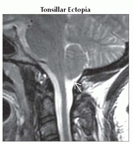 Parasagittal T2WI MR demonstrates tonsillar ectopia
 measured at 4.1 mm. Note normal rounded morphology and configuration. measured at 4.1 mm. Note normal rounded morphology and configuration.Related posts:Stay updated, free articles. Join our Telegram channel
Full access? Get Clinical Tree
 Get Clinical Tree app for offline access

|