Lumbar Radiculopathy

62 Lumbar Radiculopathy


Clinical Vignette


A 53-year-old man had a history of occasional, severe episodes of low back pain radiating down his buttock and posterior left thigh that began with an athletic injury at age 17. Typically, he experienced exacerbations every few years that lasted a few days. Precipitating factors included sitting for prolonged periods, activities such as jogging, or playing hockey. In general he “toughed out” these exacerbations by forcing himself out of bed in the morning despite the pain, and continuing with his usual activities and being careful not to suddenly bend over. If his symptoms persisted, it was necessary to use simple analgesics, low-dose muscle relaxants, and to “take it easy.” After a weekend of skiing, he developed severe left sciatica that progressively worsened over a 3-day period. The pain was excruciating, kept him up at night and did not respond to the usual medications. Getting out of bed in the morning was very painful and he had to force himself up despite the “paralyzing” pain. He noticed left foot drop with paresthesiae over his great toe. Straining or coughing further exacerbated the discomfort. He went to see a neurosurgeon; en route, routine jolting of the car significantly exacerbated the pain. Neurologic exam demonstrated a left foot drop, marked lumbosacral paravertebral muscle spasm, diminished lumbar lordosis, and an inability to tolerate straight leg raising on the left. Magnetic resonance imaging (MRI) demonstrated an extruded disc fragment at L4–5 interspace with compression of the left L5 root. A micro-hemi-laminectomy was performed, the disc fragment removed, and the nerve root decompressed. The sciatic pain was relieved the next morning.


Comment: This patient’s course was typical for an intermittent, recurrent, subacute lumbosacral radiculopathy; his intermittent symptoms had always improved with conservative therapy. The sudden onset of an acute severe radiculopathy secondary to disc extrusion with excruciating pain and the rapid development of a foot drop over a few days led to successful surgical intervention.


Lumbosacral radiculopathy, frequently called “sciatica,” is one of the most common neurologic afflictions, typically affecting 1% of the population/year. Most individuals with sciatica experience some degree of chronic low back pain. These symptoms are a major cause of disability and are the primary cause of workers’ compensation disability in the United States.


Clinical Presentation


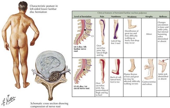
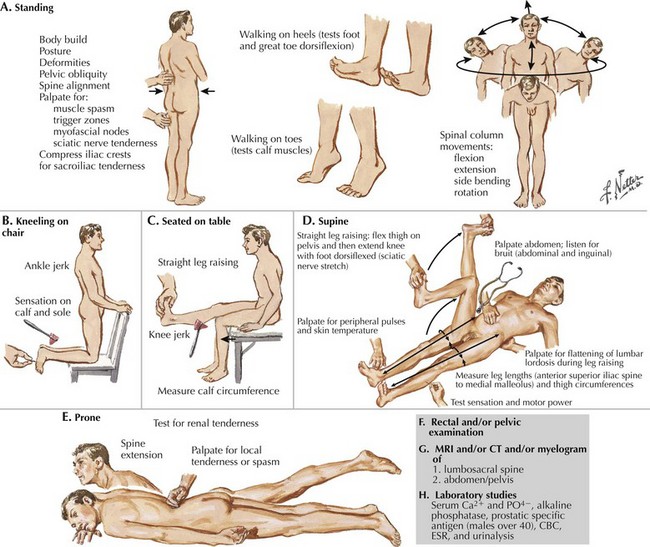
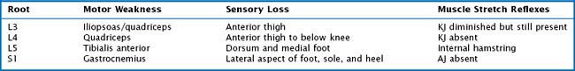
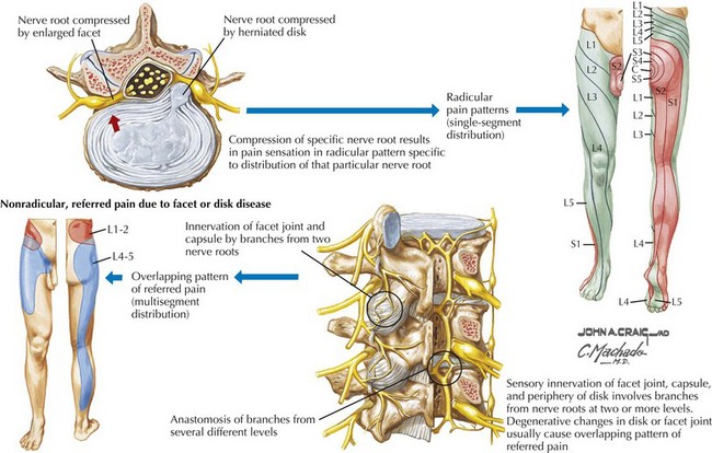
Sciatic pain may occur acutely or evolve more gradually; when the onset is sudden, it may be spontaneous or related to a specific incident, sometimes a seemingly trivial event, such as bending over to make a bed. The symptoms may be minor and clinically inconsequential or significant requiring urgent evaluation and treatment (Fig. 62-1). Depending on the specific nerve root involved, the pain may be classic sciatica with radiation down the posterior aspect of the leg into the foot, as is seen with compression of the L5 or S1 roots (Figs. 62-2 and 62-3). At higher levels, with L3 or L4 root compression, the pain may radiate to the anterior thigh. The clinical signs of lumbar radiculopathy are due to the specific level of involvement (Table 62-1), and the most common levels of nerve root irritation are L5 and S1 roots, followed less commonly by L4 and L3 roots. It is rare to have involvement of the higher roots (L1 and L2).


In the adult, the spinal cord ends between L1 and L2; therefore the nerve root compressed by disc herniation depends upon whether the lesion is medial in the spinal canal or lateral in the neural foramen. The exiting root passes around the pedicle cephalad to the disc space. Therefore a lesion occurring at the disc space in the spinal canal compresses the passing root, the root with the next lower number. For example, a medial disc rupture in the spinal canal at L4–5 will compress the L5 root, whereas less commonly the disc rupturing laterally in the neural foramen will compress the L4 root.


Etiology


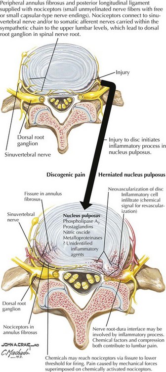
The most frequent cause of lumbar radiculopathy is a herniated lumbar disc, due to herniation of the nucleus pulposus, usually occurring with an equal frequency at the lowest two levels, L4–5 and L5–S1 (Figs. 62-4 and 62-5; see Fig. 62-1). Only ~5% of lumbar disc herniations occur at higher levels. Herniation is the last manifestation of disc degeneration that is an ongoing process in all humans. Hence, disc herniation is uncommon in youth, although occasionally teenagers and rarely toddlers have symptomatic herniations. Disc herniation occasionally occurs with spinal stenosis and may be the cause of rapid deterioration. Most lumbar radiculopathies are unilateral; bilateral sciatica has an ominous significance, suggesting compression of the cauda equina; these patients are at risk for loss of sphincter functions as well as sexual function in males. Early recognition is essential, as even after expeditious decompression, sphincter control and potency may not always return. Rarely spondylosis with foraminal encroachment resulting from disc degeneration may cause radiculopathy.


Stay updated, free articles. Join our Telegram channel

Jun 4, 2016 | Posted by in NEUROLOGY | Comments Off on Lumbar Radiculopathy

Full access? Get Clinical Tree

Get Clinical Tree app for offline access