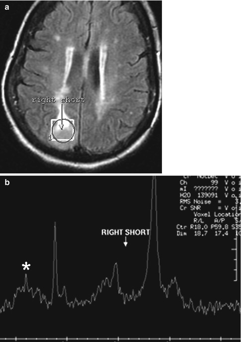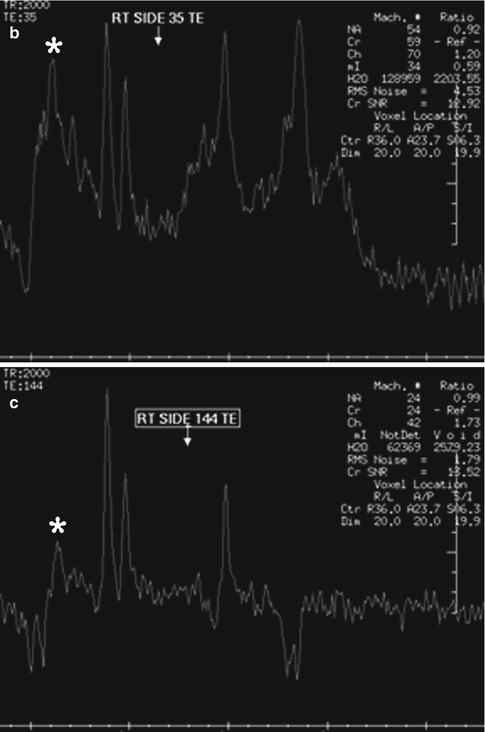
Fig. 37.1
Mannitol. The patient with a right frontal glioblastoma received mannitol. Intratumoral and peritumoral single-voxel spectroscopy (a) of the right frontal lobe(square region of interest) performed at short TE of 35 ms (b) and at intermediate TE of 144 ms (c) show a peak centered at 3.8 ppm
37.4 Differential Diagnosis
A broad peak on MRS between 3.6 and 3.8 ppm can also be found in the peritumoral edema associated with glioblastoma (Fig. 37.2), which is attributable to the α-hydrogens of amino acids, such as Glx, myo-inositol, and other metabolites. A peak at 3.8 ppm can be observed at low TE MRS in meningiomas (Fig. 37.3), which can help differentiate these from other brain tumors.
Fig. 37.2
Peritumoral edema. Single-voxel MRS of the right temporo-occipital glioblastoma shows metabolite peaks in the 3.6–3.8 range