Metabolic Disorders


Metabolic Disorders


Patricia L. Musolino

Katherine B. Sims


INHERITED METABOLIC DISEASES WITH NEUROLOGICAL INVOLVEMENT

This section is designed to allow the reader to quickly screen through the main metabolic disorders w/ neurological si/sx presenting during infancy or childhood. The first subsection presents diagnostic algorithms. The subsequent subsection presents more detailed information about specific disorders. A current expert review for each disease is referenced from the NCBI/NIH GeneReviews chapter where their up-to-date contact information is also available. Detailed instructions on diagnostic testing can be found through www.genereviews.org (see Tables 7.1, 7.2 and 7.3, Figures 7.1, 7.2 and 7.3, and Table 7.4).


TABLE 7.1 Most Common Presentations of Neurologic Metabolic Diseases by Age


Age


Neonatal 1-28 d


Late Infancy 1-12 mo


Childhood


Adolescence/Adulthood


Neuro si/sx


lethargy, diff feeding, hypotonia, irritability, seizures


lethargy or coma w/ nonfocal neuro exam a/w fasting


psych d/o, ataxia, movement d/o, recurrent episodes of coma, paraplegia, seizures, visual d/o


movement d/o, psych d/o, ataxia, peripheral neuropathy, seizures, recurrent episodes of confusion


Other si/sx


Vomiting, abnl respiration, metab acidosis, urine maple syrup smell (MSUD), sweaty foot odor ( isovaleric acidemia, glutaric aciduria type II)


recurrent vomiting, liver dysfunction, metabolic acidosis


vomiting, bone deformities, coarse facies, hepatosplenomegaly


recurrent rhabdomyolysis, myoglobinuria, cardiomyopathy


DDx


UCD, OTC, aminoacidopathies, MSUD, organic acidurias, fatty acid oxidation defects, medium chain ACoA dehydrogenase deficiency (MCAD)


same as for neonatal +: peroxisomal and lysosomal disease, Niemann-Pick, l-cell disease, MPS VII, alpha-1-antitrypsin, hemochromatosis


lysosomal d/o: Gaucher III, NCLs, Niemann-Pick, RCD


Movement d/o: metal metab d/o, RCD, neurotransmitter disorders, intermediary metab d/o


Peripheral Neuropathy. porphyrias, Refsum, PDH, CT×, Wilson, Fabry, ALD, RCD.


Spastic paraparesis: UCD, homocysteine remethylation defects, biotinidase def, PKU, NKH, DRD, cerebral folate def, CT×, ALD, lysosomal, Sjögren-Larsson


Recurrent confusion: UCD, porphyria, homocysteine remethylation disorders, CTX


Chronic psych sx. homocystinuria, Wilson disease, lysosomal d/o, NCLs, NKH, MPS III


Adapted from Paritosh P. Pocket Pediatrics. Philadelphia, PA: Lippincott Williams & Wilkins; 2009.


TABLE 7.2 Basic Initial Workup for Suspected Metabolic Disorder


Test/Study


Abnormalities of Interest


Urine: collect each fresh sample separately in clean container and stored at 4°C (short term). Freeze at-20°C. Compare samples collected before and after Rx.


Smell (special odor), look (special color) Acetone


Reducing substances


Keto acids (DNPH)


pH


Sulfitest


Sulfite oxidase


Electrolytes (Na+, K+, phos)


Uric acid (search for hyperuricuria)


Organic acids (quantitative)


Blood: 5 mL plasma (heparinized) at-20°C and10 mL whole blood in EDTA (for molecular DNA studies) and blood on filter paper(“Guthrie test”)


Blood cell count


Electrolytes (look for anion gap, glucose, calcium)


Blood gas (pH, pCO2, HCO3”, pū2)


Uric acid


Prothrombin time


Transaminases (and other liver tests)


Plasma ammonia (on ice)


Lactic acid (on ice; STAT processing)


Pyruvic acid (requires special de-proteinization tube)


3-OH-butyrate, acetoacetate (ketones)


Free fatty acids


Serum amino acids (quantitative)


CSF: neurotransmitters (collected in specific tubes & transported in dry ice); collect 2 mL STAT (on ice) for chemistries & lactic acid


Opening and closing pressures


Cell count, protein, glucose (obtain simultaneous serum glucose)


Lactic acid (on ice)


Pyruvate


Ammonia


Amino acids


Neurotransmitters, neopterins, biopterins, MTHF


ECHO/EKG


Cardiomyopathy in storage diseases


CXR/KUB


Organomegaly


Nephrocalcinosis


Adapted from Saudubray JM, Charpentier C. Chapter 66. In: Scriver CR, et al. (eds). The Metabolic and Molecular Bases of Inherited Diseases. 8th ed. New York, NY: McGraw-Hill; 2000, with permission.


TABLE 7.3 Advanced Workup for Suspected Metabolic Disorder


Study


Abnormalities of Interest


MRI/MRS brain


T1, T2 abnormalities; lactate, NAA, creatine peak analysis


Skin biopsy (fibroblast culture)


Enzyme analysis in cultured fibroblasts (FAO, PDH/PC) Staining for lipids, glycogen, cholesterol, fatty acids NP-C (cholesterol esterification, filipin staining) DNA analysis


EM for storage inclusions (lysosomal, mitochondrial)


Muscle biopsy


Light & electron microscopy, immunohistochemistry, trichrome for ragged red fibers (RRF, mitochondrial diseases), lipid storage


Different patterns of myotubular organization in myopathies


Liver, muscle biopsy (fresh & frozen at -70)


Electron transport chain (ETC activity), coenzymeQ10, acylcarnitines & carnitines (free, total, esterified)


Staining for accumulation of lipids, glycogen, cholesterol, fatty acids; immunohistochemistry


EMG/SSEP


Lower motor neuron disease pattern


Neuropathies


Myopathies


Brain US (neonates)


White matter changes


Ventriculomegaly


Intraventricular hemorrhage (IVH)


Neonatal strokes


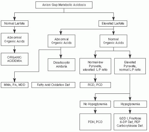
FIGURE 7.1 Metabolic Emergencies: Anion Gap Metabolic Acidosis. Adapted from Saudubray JM, Charpentier C. In: Scriver CR, et al. (eds). The Metabolic and Molecular Bases of Inherited Diseases. 8th ed. New York, NY: McGraw-Hill; 2000 and Fenichel GM. Clinical Pediatric Neurology. 6th ed. Philadelphia, PA: Saunders, Elsevier; 2009.


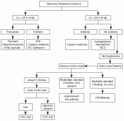
FIGURE 7.2 Metabolic Emergencies: Neonatal Hyperammonemia. Adapted from Saudubray JM, Charpentier C. In: Scriver CR, et al. (eds). The Metabolic and Molecular Bases of Inherited Diseases. 8th ed. New York, NY: McGraw-Hill; 2000 and Fenichel GM. Clinical Pediatric Neurology. 6th ed. Philadephia, PA: Saunders, Elsevier; 2009.


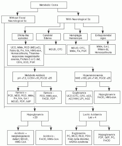
FIGURE 7.3 Metabolic Emergencies: Metabolic Coma. Adapted from Saudubray JM, Charpentier C. In: Scriver CR, et al. (eds). The Metabolic and Molecular Bases of Inherited Diseases. 8th ed. New York, NY: McGraw-Hill; 2000 and Fenichel GM. Clinical Pediatric Neurology. 6th ed. Philadelphia, PA: Saunders, Elsevier; 2009.


TABLE 7.4 Differential Diagnosis of Lactic Acidosis


Erroneous elevation


Poor collection technique (use of a tourniquet, slow blood flow), poor sample handling (not on ice or processing delay), physiologic, anaerobic exercise


Systemic diseases


Hypoxia, hypotension, shock, sepsis, renal failure, cardiac failure/cardiomyopathy, short bowel syndrome (D-lactate)


Cerebral diseases (can cause ↑ CSF lactate levels)


Prolonged seizures, meningitis/encephalitis, cerebral ischemia, malignancy, other metabolic disorders


Metabolic diseases


Amino acid disorders, organic acidurias, urea cycle defects, pyruvate metabolism disorders, Krebs cycle defects, mitochondrial OXPHOS disorders, fatty acid oxidation disorders, disorders of liver glycogen metabolism, disorders of liver gluconeogenesis, biotinidase deficiency


Other


Thiamine deficiency


Toxin exposure (carbon monoxide, methanol)


Management of Metabolic Emergencies

When suspected metabolic disorder or unknown source of metabolic acidosis, hyperammonemia, or coma, Start Rx Before confirmation of diagnosis.

1. STOP protein, fat, galactose, & fructose

2. Consult metabolic specialist

3. Remove metabolites


  • Hyperammonemia: immediate hemodialysis (HD) for coma, ventilator dependency, or signs of cerebral edema


  • Acidosis: give bicarbonate w/ frequent ABGs, severely acidotic → HD


  • Organic acidemia: Vitamin B12 (1 mg) IM for B12 responsive form of methylmalonic acidemia. Biotin (10 mg) PO or NGT for biotin-responsive carboxylase deficiency. May need HD or peritoneal dialysis if coma


  • Urea cycle defect: 6 cc/kg of 10% arginine HCL IV over 90 min


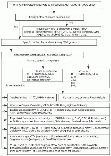
FIGURE 7.4 Progressive Spastic Paraparesis Associated with Metabolic Disorders. Adapted from Sedel F, Fontaine B, Saudubray JM, et al. Hereditary spastic paraparesis in adults associated with inborn errors of metabolism: a diagnostic approach. J Inherit Metab Dis. 2007;30(6):855-864, with permission.

4. Prevent catabolism


  • IV glucose (calories needed to keep euglycemia) D10 150 cc/kg/d w/ electrolytes


  • STOP protein (after 24-48 h restart essential amino acids), IV lipids should be given for urea cycle defects


Diagnostic Algorithms by Main Presenting Symptoms


Progressive Spastic Paraplegia


Ataxia

See Figures 7.5 and 7.6.

Epilepsies Associated with Metabolic Diseases


Movement Disorders and Extrapyramidal Signs

See Figures 7.8 and 7.9.

Peripheral Neuropathy Secondary to Inborn Error of Metabolism


Ocular Findings in Inborn Errors of Metabolism


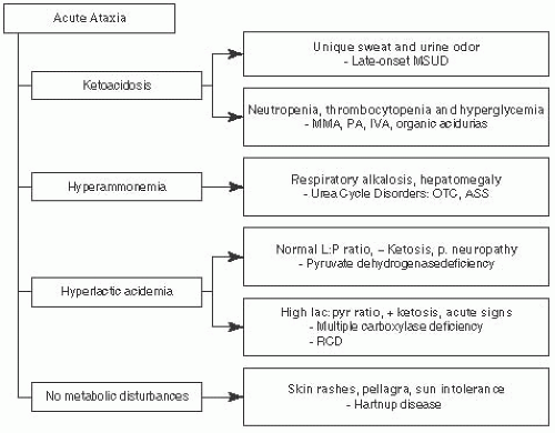
FIGURE 7.5 Acute Ataxia Associated with Metabolic Disorders. Adapted from Saudubray JM, Charpentier C. In: Scriver CR, et al. (eds). The Metabolic and Molecular Bases of Inherited Diseases. 8th ed. New York, NY: McGraw-Hill; 2000, with permission.


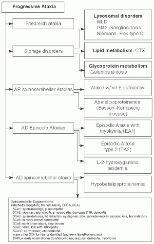
FIGURE 7.6 Progressive Ataxia Associated with Metabolic Disorders. Adapted from Saudubray JM, Charpentier C. In: Scriver CR, et al. (eds). The Metabolic and Molecular Bases of Inherited Diseases. 8th ed. New York, NY: McGraw-Hill; 2000, with permission.


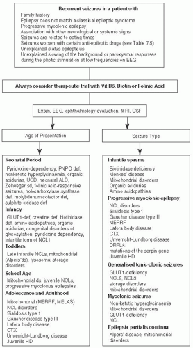
FIGURE 7.7 Epilepsies Associated with Metabolic Disorders. Adapted from Sedel F, Fontaine B, Saudubray JM, et al. Hereditary spastic paraparesis in adults associated with inborn errors of metabolism: a diagnostic approach. J Inherit Metab Dis. 2007;30(6):855-86450 and Wolf NC. Epilepsy in inborn errors of metabolism. Epileptic Disord. 2005; 7 (2): 67-81, with permission.51


TABLE 7.5 Anticonvulsants to be Avoided in Metabolic Disorders


Disease


Drugs that may Exacerbate Epilepsy or Metabolic Attacks


PME


Phenytoin, carbamazepine, gabapentin, vigabatrin, tiagabine, lamotrigine


GLUT-1 deficiency


Diazepam, phenobarbital, oxcarbamazepine


Porphyrias


Valproate, lamotrigine, oxcarbamazepine, carbamazepine, phenytoin, topiramate


Urea cycle defects


Valproate


Mitochondrial disorders


Valproate


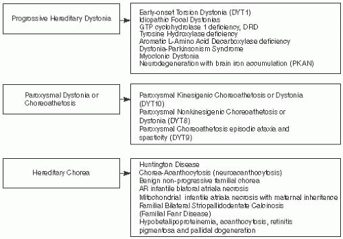
FIGURE 7.8 Movement Abnormalities Associated with Metabolic Disorders.


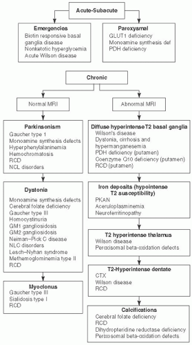
FIGURE 7.9 Movement Disorders Associated with Metabolic Disorders, Characterized by Onset and MRI Findings. Adapted from Sedel F, Saudubray JM, Roze E, et al. Movement disorders and inborn errors of metabolism in adults: a diagnostic approach. J Inherit Metab Dis. 2008;31(3):308-318, with permission.50


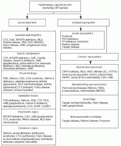
FIGURE 7.10 Peripheral Neuropathies Associated with Metabolic Disorders. Adapted from Sedel F, Baumann N, Turpin JC, et al. Psychiatric manifestations revealing inborn errors of metabolism in adolescents and adults. J Inherit Metab Dis. 2007;30(5):631-641, with permission.


TABLE 7.6 Ocular Findings Associated with Metabolic Disorders


Disorder


Optic glioma


NF type I


Retinitis pigmentosa


Peroxisomal disorders


Sjögren-Larsson syndrome (ichthyosis, spastic paraplegia, MR)


NCL disorders


Refsum disease


Kearns-Sayre syndrome


Pantothenate kinase deficiency/infantile neuroaxonal dystrophy


Bardet-Biedl syndrome (obesity, polydactyly, MR)


Usher type II (deafness, severe MR)


Joubert (MR, vermis atrophy, hyperventilation episodes)


NARP


MILS


Abetalipoproteinemia


Congenital defects of glycosylation


RCD


LCHAD


Mucopolysaccharidoses type I, II, III, IV


Macular degeneration (cherry-red spots)


MLD


Niemann-Pick disease


Wolman disease


Galactosialidosis


GM1 gangliosidosis


GM2 gangliosidosis


Optic atrophy or central blindness


ALD


MLD


Krabbe disease


Zellweger spectrum


Refsum disease


MELAS


LHON


Canavan disease


3-methylglutaconic aciduria


NCL disorders


Homocystinuria


Lens dislocation


Sulfite oxidase deficiency


Molybdenum cofactor deficiency


Homocystinuria


Marfan syndrome


Ehlers-Danlos syndrome


Neonatal cataracts


Galactosemia


Galactokinase deficiency


Tyrosinemia type II


Peroxisomal disorders


Glucose-6-phosphate dehydrogenase deficiency


Sorbitol dehydrogenase deficiency


Lowe syndrome


Cockayne syndrome


Congenital infection (syphilis, rubella, mumps)


Infantile cataracts


RCD


Sialidosis


α-mannosidosis


Mevalonic aciduria


Adapted from Saudubray JM, Charpentier C. In: Scriver CR, et al. (eds). The Metabolic and Molecular Bases of Inherited Diseases. 8th ed. New York, NY: McGraw-Hill; 2000, with permission.


DISEASES AFFECTING METABOLIC PATHWAYS


Metal Metabolic Disorders


Wilson Disease1

Familial hepatolenticular degeneration

Defect: Defect in copper-transporting ATPase 2, (ATP7B).

FHx: AR, Mediterranean descent

Presentation: Classically p/w movement d/o (tremors, poor coordination, loss fine-motor, choreathetosis) + hepatic failure. Hepatic failure predominates in children w/ onset at < 10 y. If onset in 2nd decade, one neurologic
si/sx such as gait or speech disturbance, unchanged for years & then development of movement d/o (e.g., dysarthria, dystonia, dysdiadochokinesia, facio-linguo-pharyngeal rigidity, & gait difficulties). Other sx: rest & action tremor (rubral tremor) ± chorea & parkinsonism. Psychiatric si/sxprecedes movement d/o in 20% of cases.

Other si/sx: Hepatic failure/cirrhosis, corneal Kayser-Fleischer ring (seen in 100% cases w/ CNS involvement) & azure lunulae of the fingernails. Fanconi syndrome.

Prognosis: Variable. Can have normal life span if therapy instituted early.

Dx: Imaging: MRI: cerebral atrophy, T2 hypointensity of the globus pallidus, & T2 hyperintensity of caudate, putamen, thalamus, dentate nucleus, & pons. Labs: Low serum copper & ceruloplasmin. Elevated LFTs, increased urinary copper. Liver biopsy: Elevated copper in liver w/ assay of copper content by spectrometry (copper content > 250 mcg dry weight of liver). Definitive dx: Neuro-ophthalmologic slit-lamp exam showing corneal Kayser-Fleischer rings, mutational analysis (ATP7B). Screen siblings.

Rx: Lifelong; copper-chelating agents: D-penicillamine, trientine, tetrathiomolybdate; zinc, antioxidants, restriction of foods very high in copper. Liver transplant controversial.


Aceruloplasminemia2

Defect: Mutation of the ceruloplasmin gene (CP) on chromosome 3q25. Accumulation of iron in liver, pancreas, brain, & retina.


Presentation: Very rare, p/w progressive neurodegeneration resulting in dystonia or chorea, dementia, retinal degeneration, & cerebellar ataxia.

Other si/sx: Microcytic anemia, diabetes.

Dx: Imaging: MRI: iron accumulation (T2 hypointensity) in the putamen, caudate, thalamus, dentate nuclei. Labs: Undetectable serum ceruloplasmin, ↓ iron, ↓ copper, ↑ ferritin, microcytic anemia. LFTs normal. Biopsy: liver iron deposits in hepatocytes & RE system. Definitive dx: DNA mutation analysis (CP).

Rx: Iron chelators (e.g., desferrioxamine, FFP), antioxidants. Avoid iron supplements.


Hemochromatosis3

Prevalence: High carrier rate (1:250), but very low incidence (phenotypic) in children.

Defect: Classic type I: C282Y gene. Class II juvenile: AR hemojuvelin gene (HFE2) and hepcidin antimicrobial peptide (HAMP). Class III: AR transferrin receptor-2 (TFR2). Class IV: AD ferroportin (FPN1).

FHx: All AR except Class IV, which is AD.

Presentation: More common in adults, but can be seen as early as 2 y (in children: cardiomyopathy & gonadal failure). Neurologic signs include: parkinsonism, cerebellar ataxia, dementia, myoclonus, action tremor, cervical dystonia.

Other si/sx: Cirrhosis & primary hepatocellular carcinoma, arthritis, DM, bronze skin pigmentation.


Prognosis: Depends on progression of disease before therapy. Children often die of cardiac cause.

Dx: Imaging: Normal MRI (may have brain atrophy). Labs: High serum iron, transferrin saturation, & ferritin. Biopsy: Hepatic iron is the most sensitive index of preclinical disease. Confirmed by hepatic iron index score >1.9 or hepatic iron >80 µmol/g dry weight of liver or grade 3 or 4 on Perls stain. Definitive dx: Liver biopsy & genetic testing.

Rx: Phlebotomy for ferritin >300 mcg/L in men; ferritin > 200 mcg/L in women, regardless of the presence or absence of symptoms.


Neurodegeneration with Brain Iron Accumulation (NBIA)4


Infantile Neuronal Dystrophy (INAD; Seitelberger disease)

FHx: AR; PLA2G6 mutations.

Presentation: Progressive degeneration of CNS and PNS. Infantile hypotonia followed by spasticity, opisthotonic posturing, optic atrophy. Death usually by 5 y.

Pathology: Axonal spheroids on skin or nerve biopsy.

Dx: MRI: diffuse cerebellar hyperintensity and atrophy ± iron in basal ganglia. EMG: anterior horn cell dysfunction, denervation. NCλζ normal; ERG, normal.

Rx: Symptomatic


Pantothenate Kinase-Associated Neurodegeneration (PKAN)

Defect: Pantothenate kinase deficiency, PANK2 mutations.


Presentation: Onset 7 to 10 y w/ dystonia, dysphagia, parkinsonism, choreathetosis, and retinitis pigmentosa. Cognitive and psychiatric disorders, along w/ spasticity follow.

Prognosis: Spastic quadriplegia and death within 5 to 10 y.

Dx: Imaging: T2 hyperintensity w/ surrounding hypointensity in medial globus pallidius (“eye of the tiger sign”). Definitive dx: PANK2 mutation analysis.

Rx: Symptomatic, deep brain stimulation for dystonia.


Menkes: Trichopolydystrophy5

Defect: Defect in the intestinal transport of copper, ATP7A gene mutation.

FHx: X-linked recessive.

Presentation: Symptoms result from secondary deficiency of copper-dependent enzymes (cytochrome-c-oxidase, dopamine-beta-hydroxylase, lysyl oxidase, etc.). Most marked are neurodegenerative changes and connective tissue abnormalities. Infants are usually hypotonic, progressing to spastic quadriparesis. Autonomic dysfunction, temperature instability, and hypoglycemia are seen in neonates.

Other si/sx: Sparse, poorly pigmented, pili torti (kinky hair), osteogenesis imperfecta, high arched palate, micrognathia, fullness of the cheeks, occipital horns.

Prognosis: Progressive encephalopathy leaves patients vegetative by 1 y, death usually by 1.5 to 2 y, no therapy. Cases surviving up to 20 y have been described.


Dx: Labs: Low plasma ceruloplasmin; copper microscopic examination of the hair (pili torti seen in carriers). Biopsy: Skin for enzyme assay in cultured fibroblasts. Definitive dx: ATP7A mutation analysis. Prenatal testing.

Rx: subcutaneous injections of copper histidine or copper chloride before 10 d of age normalizes developmental outcome.


Peroxisomal Disorders6


Refsum Disease (Phytanic Acid Oxidase Deficiency)

Defect: Defect of phytanoyl-CoA hydroxylase (PAHX or PHYH gene).

FHx: AR, more common in Scandinavian, British, German, & French.

Presentation: Insidious onset in 1st through 3rd decade w/ night blindness (retinitis pigmentosa), recurrent or chronic polyneuropathy (often symmetric, distal, vibration > pain & temp), palpable nerves, cerebellar ataxia, dysmetria, dysarthria. When presenting in infants, severe hypotonia w/ absent DTRs seen. Symptoms made worse by fasting. Other si/sx: sensorineural hearing loss, cardiomyopathy, ichthyosis, pes cavus.

Prognosis: Variable, progressive w/ periods of stability & exacerbations. Death may occur from sudden cardiac event.

Dx: Imaging: MRI: symmetrical T2 hyperintensity involving the corticospinal tracts, cerebellar dentate nuclei, & corpus callosum (only seen on infantile form). Labs: ↑ plasma branched-chain FAs (mostly phytanic acid), ↑ CSF protein (1-7 g/L). Biopsy: Skin for enzyme assay in cultured fibroblasts. Definitive dx: PAHX or PHYH mutation analysis.

Rx: Dietary modification to avoid phytanic acid ± plasmapheresis.

X-linked Adrenoleukodsytrophy (XALD), see Chapter 9.

Zellweger Spectrum disorders, see Chapter 9.


Lysosomal Disorders


GM1 Gangliosidosis7 (type I: infantile, type II: late infantile/juvenile, & type III: adult)

Defect: Beta-galactosidase deficiency, GLB1 gene mutation.


Presentation: Type I (infantile onset): difficulty feeding/failure to thrive, hypotonia. Type II (late infantile/juvenile): initially ataxia, strabismus, & dysarthria, followed by seizures, mental regression, & spasticity.

Other si/sx: Type 1: hepatosplenomegaly, deafness, mental retardation, kyphoscoliosis, vertebral & hip dysplasia (resembling Hurler disease, but w/o corneal opacities & w/ macular cherry-red spots [50%]), renal disease, dilated and/or hypertrophic cardiomyopathy.

Type II: no visceromegaly & only mild skeletal abnormalities.

Prognosis: Death typically occurs within 1 y in the infantile form. Later onset associated with survival into 1st or 2nd decade.

Dx: Imaging: T2 hyperintensity of posterior putamen. Labs: Low betagalactosidase activity in leukocytes. Biopsy: Liver, heart, kidney, fibroblast show accumulation of gangliosides. Definitive dx: Beta-galactosidase activity in leukocytes & GLB1 molecular testing.

Rx: Symptomatic or Miglustat (inhibitor of glucosylceramide synthase)


GM2 Gangliosidosis, type I8: Tay-Sachs Disease (infantile, juvenile/chronic adult forms)

Defect: Hexosaminidase A deficiency; HEXA mutations.

FHx: AR, Ashkenazi Jews with high carrier rate.

Presentation: Infantile form (most common): persistent startle reaction to sound, developmental regression, followed by paralysis, dementia, & blindness. Juvenile onset (late-onset form; onset late childhood to adulthood): childhood clumsiness or incoordination, can see psychiatric presentation; associated findings include: proximal muscle weakness (typically LE

Only gold members can continue reading. Log In or Register to continue

Stay updated, free articles. Join our Telegram channel

Jun 20, 2016 | Posted by in NEUROLOGY | Comments Off on Metabolic Disorders

Full access? Get Clinical Tree

Get Clinical Tree app for offline access