Neuromuscular Disorders


Neuromuscular Disorders


Jahannaz Dastgir

Basil T. Darras


Neuromuscular disorders may present as a variety of symptoms and consult requests to the pediatric neurologist. Consults may be in reference to vague concerns regarding “floppy” infants or children with “developmental delays”. They may also be called regarding more specific complaints such as localized weakness, fatiguability, abnormal gait, myalgias or ptosis. An accurate history and a detailed exam guide testing, for example, serum/urine labs, imaging (MRI/US), electrophysiology, or biopsy (muscle/skin). Figure 6.1 illustrates the diagnostic approach to the infant with hypotonia. Differential diagnosis can be guided by the anatomy of the motor unit (Fig. 6.2).


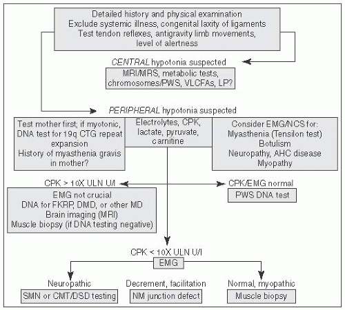
FIGURE 6.1 Approach to Hypotonia. MRI, magnetic resonance imaging; MRS, magnetic resonance spectroscopy; PWS, prader willi syndrome; VLCFA, very long chain fatty acids; LP, lumbar puncture; AHC, anterior horn cell; CPK, creatine phosphokinase; CMT, Charcot Marie Tooth; DMD, Duchenne Muscular Dystrophy; DSD, Dejerine Sottas Disease; EMG, electromyography; NCS, nerve conduction studies; NM, Neuromuscular (From Perlman J, ed. Neonatology: Questions and Controversies. London, UK: Elsevier; 2012, with permission.)


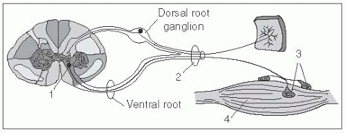
FIGURE 6.2 The Four Anatomic Stations Underlying Lower Motor Neuron Weakness. 1, anterior horn cell; 2, peripheral nerve; 3, neuromuscular junction; 4, muscle fiber (From http://www.neuroanatomy.wisc.edu/SClinic/Weakness/Weakness.htm.)


DISORDERS OF THE MOTOR NEURON


Spinal Muscular Atrophy (SMA)1

DEFINITION: Heterogenous group of disorders (Table 6.1) with progressive degeneration of spinal cord anterior horn cells ± motor nuclei in the brainstem (extraocular and sacral motor nerves and cardiac and smooth muscles are selectively spared).


TABLE 6.1 Classification of Spinal Muscular Atrophies (SMA)


SMA1/Werdnig-Hoffmann Disease (onset: <6 mo, never sit)


Genetics: AR homozygous SMN1 deletion (95%); <1-2 SMN2 copies


Exam: severe weakness of the limbs and intercostal muscles; paucity of spontaneous movement at the shoulder and hip girdle; some antigravity movements in the hands and feet. Atrophy of the tongue may be associated with fasciculations. DTRs are absent. Never sit. Normal intellect


Prognosis: >9Å% mortality; few survive to 2nd decade


SMA2 (onset: 6-18 mo, sit, but never walk)


Genetics: AR homozygous SMN1 deletion with usually 3 SMN2 copies


Exam: Lower extremities are more involved than upper extremities; associated with a tremor affecting the upper extremities (polyminimyoclonus); tongue may have fasciculations. DTRs are absent. Sit, but never walk. Normal intellect


Prognosis: variable, most survive to 2nd/3rd decade


SMA3/Kugelberg-Welander (onset >18 mo [type 3A, <3 y; type 3B, >3 y], able to walk)


Genetics: AR homozygous SMN1 deletion with 3-4 SMN2 copies


Exam: proximal symmetric weakness; muscle weakness may not manifest until adult life. Limb and tongue fasciculation. Able to walk. Type 3A: 20% walking at 40 y; type 3B: 60% walking at 40 y. Normal intellect


Prognosis: will survive into adult life


SMA4/Adult form (onset: after 20 y, walk with waddling gait)


Genetics: 30% AD; AR may be SMN-related, X-linked


Exam: proximal muscles are predominantly involved; impaired joint mobility, waddling gait, lumbar lordosis, and protuberant abdomen. ± DTRs, limb and tongue fasciculations, and tremor


Prognosis: age of symptom onset correlates with cessation of ambulation


EPIDEMIOLOGY: Incidence 1/6,000 to 10,000 live births. 2nd most common hereditary neuromuscular disease after Duchenne muscular dystrophy (DMD) and the 2nd most common AR disease in children after cystic fibrosis.

GENETICS: Due to deletions in “survival of motor neuron” (SMN) gene, of which there are 2 types: SMN1 and SMN2. Homozygous deletions (95%) of SMN1 lead to more severe forms that may vary in presentation depending on the number of SMN2 genes that are present (the more copies of SMN2, the less severe the manifestation of the disease).

CLINICAL PRESENTATION: Widespread muscular denervation and atrophy, with marked reduction of muscle power and spontaneous movement, in a symmetric and proximal > distal distribution. Diaphragm involvement depends upon type of SMA. No cognitive impairment.

DIAGNOSIS: Genetic testing, EMG (spontaneous muscle activity, fibrillation potentials, and residual motor unit potentials that are increased in amplitude and duration), muscle biopsy (not common practice at present; demonstrates small and large group atrophy); CK may range from normal to 5× ULN.

TREATMENT: Symptomatic, aimed at maintaining joint mobility and avoiding contractures. Monitor and support other potential complications, including dysphagia, scoliosis, restrictive lung disease, and poor nutrition.


Other Spinal Muscular Atrophies


(1) Juvenile Segmental SMA (Hirayama)

GENETICS: Primarily a sporadic disorder; may be related to positional cervical cord compression.

CLINICAL: Variable; tends to have asymmetric weakness and atrophy of the unilateral or bilateral hand and forearm muscles. May involve upperlimb amyotrophy ± bulbar amyotrophy, with pyramidal signs and a slowly progressive course.

PROGNOSIS: Progression for 2 to 6 y, then plateau.


(2) Scapuloperoneal Neuropathy

GENETICS: TRPV4 gene on chromosome 12q24.11 (AD)

CLINICAL PRESENTATION: Progressive weakness in the scapuloperoneal and distal muscles; may also be associated with laryngeal palsy.

PROGNOSIS: Progressive; disease is more severe in succeeding generations (anticipation).


(3) Childhood Bulbar SMA (Fazío-Londe and Brown-Vialetto-Van Laere)

GENETICS: C20orf54 gene on chromosome 20p13 (AR).

CLINICAL PRESENTATION: Bulbar palsy; Brown-Vialetto-Van Laere also associated with sensorineural deafness. Fazio-Londe may have prominent facial weakness and ophthalmoplegia.

PROGNOSIS: Onset in 1st 2 decades with variable rate of progression.


Acquired Motor Neuron Disease


(1) Acute Poliomyelitis

DEFINITION: Highly contagious via oral-oral and fecal-oral routes. In endemic areas, wild polioviruses can infect virtually the entire human population.


INCIDENCE: The most common cause of acquired anterior horn cell disease. Eradicated in the United States, but prevalent in developing countries. Poliolike diseases due to coxsackie and other echoviruses have also been described.

CLINICAL PRESENTATION: Ranges from asymptomatic to minor illness (1-5 d post exposure; fever, malaise, sore throat, GI symptom) to major illness (4-10 d post exposure; aseptic meningitis, fever, pain). Paralytic disease will manifest in 50% of those with major illness 2 to 5 d after illness. Exam shows localized fasciculations, intense myalgia, hyperesthesia; may be focal or asymmetric; legs > arms > bulbar symptoms. May also have acute dysautonomia.

DIAGNOSIS: CSF pleocytosis and IgM poliovirus specific; stool positive 90% by 20 d; EMG/NCS: selective loss of motor neurons/axons at 7 to 10 d.

TREATMENT: Some cases only involve temporary paralysis and recover. Others may be permanent and supportive measures are necessary.


(2) West Nile Encephalomyelitis

DEFINITION: Tick-borne flavivirus that primarily causes a meningoencephalitis but may also lead to a poliolike disease.

CLINICAL PRESENTATION: Fever, meningeal signs, and CSF pleocytosis that may be followed by a focal asymmetrical flaccid paralysis over the next few days.

DIAGNOSIS: CSF pleocytosis and positive for West Nile virus IgM; EMG/NCS: selective loss of motor neurons/axons at 7 to 10 d; MRI with T2 hyperintensities in the anterior horn cells.

TREATMENT: Supportive. Antiretroviral treatment is under development.


DISORDERS OF THE PERIPHERAL NERVE


Hereditary Motor and Sensory Neuropathies (HMSN)2

HMSNs account for ˜40% of childhood chronic neuropathies. They involve degeneration of myelin sheaths and disorder of axons and lead to a distal paralytic amyotrophy involving the lower > upper limbs that is also associated with areflexia. In this disease group the neuropathy is primary as opposed to only part of the disorder.3


(1) Charcot-Maríe-Tooth Disease (CMT)4

EPIDEMIOLOGY: Most common inherited neurologic condition, affecting ˜1 in 2,500 people.

GENETICS: >40 genes are known to cause CMT; the majority are AD; X-linked and AR inheritances have also been described. An exhaustive list can be found at www.genetests.org.

CLINICAL PRESENTATION: Variable in age of onset (typically 1st-2nd decade) and speed of progression. Classic phenotype involves steppage gait, pes cavus, sensory loss in a stocking/glove distribution, inverted champagne bottle legs, and atrophy in the hands (all typically symmetric). Patients have decreased or absent deep tendon reflexes, impaired proprioception, and may or may not complain of neuropathic pain. May also be a/w other features distinctive to genetic phenotype: optic atrophy (CMT2A); vocal cord and respiratory involvement (CMT2C, 4A), predominant hand weakness (CMT2D), facial/bulbar symptoms (CMT4B1), scoliosis (CMT4C), sensory ataxia (CMT4F), upper-motor-neuron-like changes (CMT4J).


DIAGNOSIS: Nerve conduction studies allow for classification into demyelinating, axonal, or intermediate groups.


(2) HMSNs Associated With Metabolic and Degenerative CNS Disorders

Abetalipoproteinemia, adrenomyeloneuropathy, amyloidosis, ataxia-telangiectasia, carbohydrate-deficient glygoprotein syndrome, cerebrotendinous xanthomatosis, Cockayne syndrome, Chédiak-Higashi disease, Fabry disease, Farber lipogranulomatosis, GM2 gangliosidosis, Krabbe disease, metachromatic leukodystrophy, mitochondrial disease (Leigh, NARP, MNGIE), neuroaxonal dystrophies, Niemann-Pick disease, porphyria, Refsum disease, Tangier disease, tyrosinemia, vitamins E and B12 deficiency.


(3) Hereditary Sensory and Autonomic Neuropathies (HSAN)5

DEFINITION: Hereditary group of disorders involving the peripheral nervous system that involve progressive degeneration of the sensory and autonomic neurons. Categorized as types I to V on the basis of age at onset, mode of inheritance, and predominant clinical feature.

GENETICS: 12 genes have been discovered to date (www.genetests.org). Patients with AD forms (HSAN I) typically show juvenile-adult onset of disease; AR forms (HSAN II-V and HSAN w/ spastic paraplegia) show an earlier onset (e.g., congenital or in childhood).

CLINICAL PRESENTATION: Loss of sensations of pain and temperature are the most common symptoms and frequently lead to sensory loss and chronic ulcerations in the feet and hands of the patient. These lesions can lead to extensive soft tissue infections or osteomyelitis and subsequently amputation of the affected limb.

DIAGNOSIS: EMG/NCS show axonal damage to sensory neurons, but demyelination might additionally be present.


Immune-Mediated/Inflammatory Neuropathies


(1) Acute Inflammatory Demyelinating Polyneuropathy (AIDP), or Guillain-Barré Syndrome (GBS)6,7

DEFINITION: Autoimmune neuromuscular disorder with loss of immunologic tolerance and autoreactive T lymphocytes, with antibodies and complement damage to myelinated peripheral nerves.

INCIDENCE: 0.6 to 4 cases per 100,000; men > women; incidence increases with age. Uncommon in the 1st few years of life, but rare neonatal cases have been reported.

CLINICAL PRESENTATION: 2/3 of cases with an antecedent vaccination or infection within 6 wk prior to symptoms onset (e.g., EBV, Mycoplasma pneumoniae, Campylobacter jejuni, and CMV). Progressive, symmetrical muscle weakness and diminished or absent deep tendon reflexes. Around 50% to 80% (particularly younger children) complain of pain in the back, buttock, or limb and refuse to walk. Symptoms tend to ascend from lower to upper limbs (but not in all cases); weakness of facial muscles in 50%; weakness peaks by 2 to 4 wk post symptoms onset and is followed by progressive recovery. Monitor for respiratory failure (˜17%) and dysautonomia (cardiac arrhythmia, hypertension, hypotension, ileus, urinary retention, and difficulty with thermoregulation, in 20%-40%). Miller Fisher variant (much more rare) is also associated with the triad of external ophthalmoplegia (may be asymmetric), ataxia, and areflexia, but also ptosis, mydriasis, facial and oropharyngeal weakness, myalgias, and paresthesias.


DIAGNOSTIC WORKUP: (1) CSF: elevated protein in ˜80% (may be normal in 1st wk of illness), and absence of pleocytosis is known as albuminocytologic dissociation. (2) EMG/NCS: normal or decreased motor amplitudes and slowing of conduction velocities (changes may be absent when studying patients early in the disease course). (3) MRI of the brain (with gadolinium) may show cranial nerve enhancement, and of spine may show spinal root enhancement. (4) Antibodies: Miller Fischer variant is often associated with IgG reactivity to GQ1b or GT1b gangliosides.

MANAGEMENT: Close monitoring of respiratory function via serial pulmonary function tests (vital capacity) and bedside negative inspiratory flow and of cardiac function via electrocardiography and monitor. Intravenous immunoglobulin (IVIG) or plasmapheresis within the 1st 4 wk of symptom onset. Oral corticosteroids alone tend to be ineffective, but there are some reports of combination therapy with IV corticosteroids and IVIG accelerating recovery. Physical therapy is essential in the rehabilitation process.

PROGNOSIS: The disease reaches a nadir at 2 to 4 wk, with most patients recovering, but 10% to 20% are left with motor deficits of variable severity and a small fraction of patients die from complications. Adverse prognostic indicators include need for artificial ventilation throughout the course of illness, rapid onset of disease, established history of infection with C. jejuni or cytomegalovirus, and evidence of axonal loss on neurophysiologic testing.


(2) Chronic Inflammatory Demyelinating Polyneuropathy (CIDP)6

DEFINITION: Distinguished from GBS by the chronic nature of the disease.

CLINICAL PRESENTATION: Progressive or relapsing sensorimotor polyneuropathy disorder polyradiculopathy for at least 2 mo. The majority exhibit an insidious onset.

DIAGNOSIS: Diffuse nerve root thickening and gadolinium enhancement on spine MRI (may also be seen in CMT patients); elevated protein in CSF (may also be seen in CMT patients), and EMG/NCS showing normal or decreased motor amplitudes, slowing of conduction velocities, and occasionally conduction block.

TREATMENT: IVIG, plasmapheresis, corticosteroids, azathioprine (steroid sparing), and physical therapy.


(3) Acute Motor Axonal Neuropathies

DEFINTION: Acute motor axonal neuropathy (AMAN) and acute motorsensory axonal neuropathy (AMSAN) are neuropathies thought to be related to disruption of node of Ranvier function as brought about by antibodies to GM1 and GD1a.

INCIDENCE: Most commonly found in Japan, China, Mexico, and 3rd world countries; children > adults; no gender predilection, presence of seasonal peaks.

CLINICAL PRESENTATION: Prodrome includes gastrointestinal diarrhea (C. jejuni in most), an upper respiratory infection (e.g., H. influenza infection), or immunization. AMAN: ascending, symmetric paralysis of the extremities, loss of deep tendon reflexes, and variable involvement of bulbar, respiratory, ocular, and facial muscles. No complaints of paresthesias. AMSAN: will also include sensory symptoms.

DIAGNOSTIC WORKUP: (1) CSF: (AMAN) increased protein; (2) EMG/NCS: (AMAN) normal sensory testing, abnormal motor testing (seen in
first 2 wk); (AMSAN) mildly reduced nerve conduction velocities combined with a marked reduction in motor and sensory action potential amplitudes; (3) serum: (AMAN) IgG reactivity to GM1, GD1a, and GD1b positive, GQ1b negative.

TREATMENT: IVIG, plasmapheresis; corticosteroids are not recommended. AMAN has a shorter course than does GBS.


Vasculitic Neuropathies

To be considered in patients with vasculitides such as SLE, RA, and PAN.


Neuropathies Related to Infection

1. Leprosy: Myobacterium leprae; HLA-linked genes control susceptibility; predilection for superficial nerves (may see nerve enlargement), skin, anterior third of eye, upper respiratory tract, and testes. May start as a purely sensory polyneuritis predominantly affecting the cooler areas of the body and eventually leading to paralysis. Skin biopsies and serum testing aid in the diagnosis. Treatment as per current WHO recommendations.8

2. Lyme: With acute, disseminated disease, patients may develop neurologic manifestations, which can include a distal sensorimotor neuropathy or carpal tunnel syndrome. EMG/NCS shows axonal loss. Nerve biopsy shows perivascular inflammation. CSF shows mononuclear pleocytosis and moderately elevated protein. Serum ELISA is done with Western blot confirmation. With neurologic symptoms, patients should receive IV ceftriaxone therapy for 2 to 4 wk as per current recommendations.9

3. HIV: Sensory > motor neuropathy; increased frequency with higher viral load. Symptoms include pain and dysesthesias. Mainly in the feet. EMG/NCS shows distal axonal loss with or without demyelination. Many also lead to dysautonomia, multiple mononeuropathy, mononeuritis multiplex. CSF shows pleocystosis, low glucose, elevated protein.


Toxic Neuropathies

Can occur with amiodarone, amitryptyline, amphotericin, arsenic, carbon monoxide, chlorambucil, ciguatera cisplatin, cyanate, diphtheria, ethambutol, ethionamide, hydroxyquinolines, isoniazid, lead, lithium, mercury, metronidazole, n-hexane nitrofurantoin, organophosphate organic chemicals, phenytoin, thalidomide, thallium, triorthocresylphosphate, vincristine.


Localized Peripheral Nerve Disorders


Brachial Plexus Palsy

DEFINITION: May be obstetrical (caused by forces-generated labor stretching the brachial plexus beyond its resistance; most common), familial congenital, or due to intrauterine maladaption or maternal uterine malformation, congenital varicella syndrome, osteomyelitis involving the proximal head of the humerus or cervical vertebral bodies, exostosis of the 1st rib, and tumors or hemangiomas in the region of the brachial plexus.

INCIDENCE: 0.5 to 1.9/1,000 live births

CLINICAL PRESENTATION: Erb palsy is the most common manifestation (related to disruption of the upper brachial plexus at C5 and 6 nerve roots) and involves an asymmetric Moro reflex, weakness of abduction
and external rotation at the shoulder, elbow flexion, supination at the wrist, and extension at the fingers (aka “waiter’s tip”). Klumpke palsy is less common (related to disruption at the C7, C8, and T1 nerve roots) and involves weakness of the intrinsic muscles of the hand and flexors of the wrist and fingers (aka “claw hand”). A supinated forearm with absent grasp reflex may be noted on exam. Involvement of the 1st thoracic root can result in an ipsilateral Horner syndrome. The phrenic nerve (arising from C3, C4, and C5) may also be affected and lead to ipsilateral diaphragmatic paralysis.

DIAGNOSIS: Observation of arm weakness distribution. X-ray of chest, clavicle, humerus. MRI of brachial plexus. EMG to evaluate for signs of denervation.

Jun 20, 2016 | Posted by in NEUROLOGY | Comments Off on Neuromuscular Disorders

Full access? Get Clinical Tree

Get Clinical Tree app for offline access