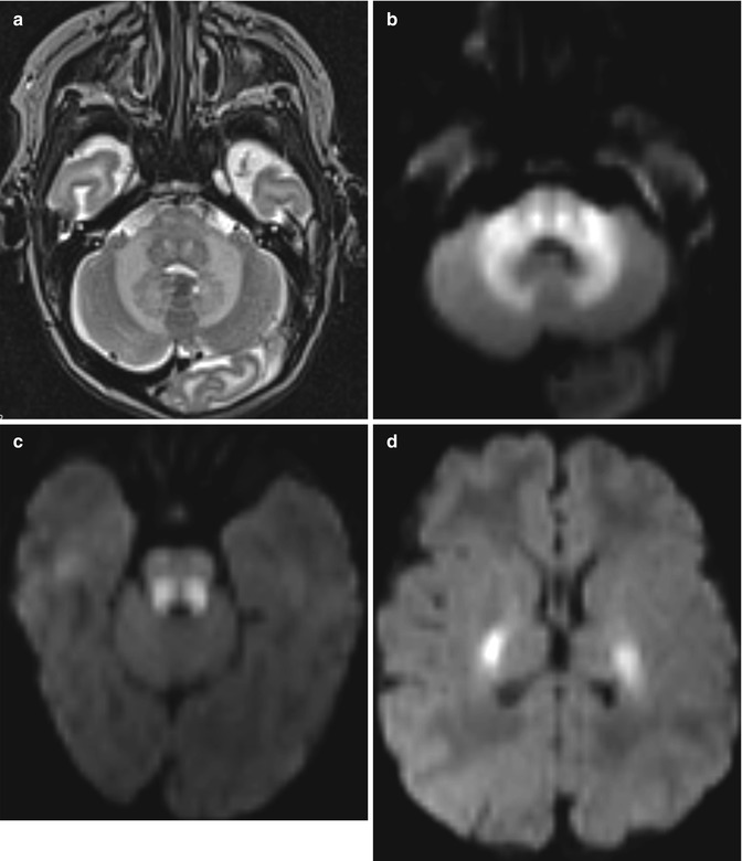
Fig. 25.1
Metronidazole-induced encephalopathy. FLAIR MR images (a–c) at the time of presentation show bilateral symmetric high signal within the dentate nuclei, red nuclei, cerebral peduncles, corpus callosum, and periventricular and subcortical white matter. An MRI obtained 2 months later (not shown) demonstrated decrease in the signal abnormalities
25.3 Discussion
Metronidazole toxicity can produce a characteristic encephalopathy that manifests as ataxia, peripheral neuropathy, and seizures. Affected patients may have normal or toxic serum concentrations. On MRI, high T2 signal in the dentate nuclei is the earliest and most common finding. This initial finding is followed by involvement of the tectum, red nucleus, periaqueductal gray matter, and dorsal pons. The dorsal medulla and the corpus callosum are less commonly involved. Lesions are often bilateral and symmetric, and the splenium is affected in all cases in which the corpus callosum is involved. The lesions are usually characterized by elevated ADC, consistent with vasogenic edema. The lesions do not enhance and do not have significant mass effect. Involvement of supratentorial structures appears to correlate with the severity of toxicity. The abnormal findings on MRI and clinical symptoms typically improve within 8 weeks following discontinuation of metronidazole.
25.4 Differential Diagnosis
The main differential consideration for metronidazole-induced encephalopathy is acute Wernicke’s encephalopathy associated with alcohol abuse. Wernicke’s encephalopathy is characterized by bilateral symmetric T2 hyperintense lesions in the regions of the mammillary bodies, medial thalami, floor of the third ventricle, periaqueductal gray matter, and tectum of the midbrain.
Other imaging differential diagnoses to consider for metronidazole-induced encephalopathy depend on the particular anatomic sites of involvement:
Brainstem: Osmotic demyelination involves the basis pontis (refer to Chaps. 2 and 36), while metronidazole-induced encephalopathy affects the dorsal pons and nuclear structures.
Corpus callosum splenium: Various demyelinating diseases, such as Marchiafava-Bignami disease (refer to Chap. 2), multiple sclerosis, and Susac’s disease (refer to Chap. 27); antiepileptic drugs (refer to Chap. 27); acute infectious encephalitis, such as influenza, Escherichia coli, mumps, adenovirus, Epstein-Barr virus, and Rotavirus; and acute toxic encephalopathy, such as from methotrexate (refer to Chap. 19) and 5-fluorouracil (refer to Chap. 20), can produce similar findings (Fig. 25.2).


