• Rib exposure. The anterior and posterior margins of the vertebral body to be resected are marked using fluoroscopy. The overlying rib is subperiosteally exposed.

• The inferior portion of the rib is subperiosteally released from the neurovascular bundle.
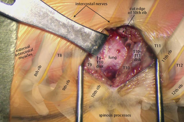
• Approximately two centimeters of rib have been resected. The pleura of the lung is visualized.
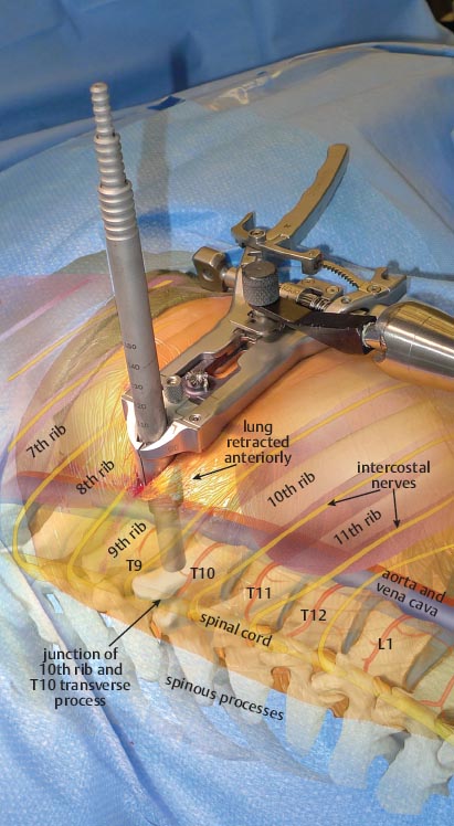
• A series of tubular dilators are placed into the defect, sweeping the lung and pleura anteriorly and resulting in an extrapleural exposure of the vertebral body.

Related posts:

Stay updated, free articles. Join our Telegram channel

Full access? Get Clinical Tree


