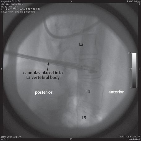
• Biplanar fluoroscopy is used throughout the procedure.
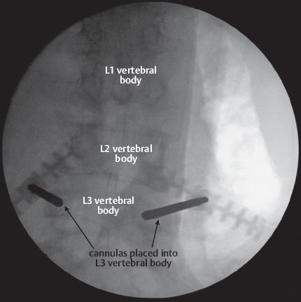
• The starting position for the cannula should be at the 10 o’clock or 2 o’clock position of the pedicle (superior-lateral corner depending upon the left or right side of the spine). This position places the cannula the farthest from the exiting nerve root. The cannula is advanced such that on the AP side it never crosses the medial edge of the pedicle until it crosses the posterior verterbral body line on the lateral image.
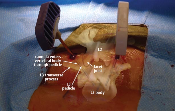
• The cannula is started lateral to the facet joint, thereby avoiding any damage to the facet capsule.
Related posts:

Stay updated, free articles. Join our Telegram channel

Full access? Get Clinical Tree


