66 Mononeuropathies of the Lower Extremities
Sciatic Neuropathies
Clinical Vignette
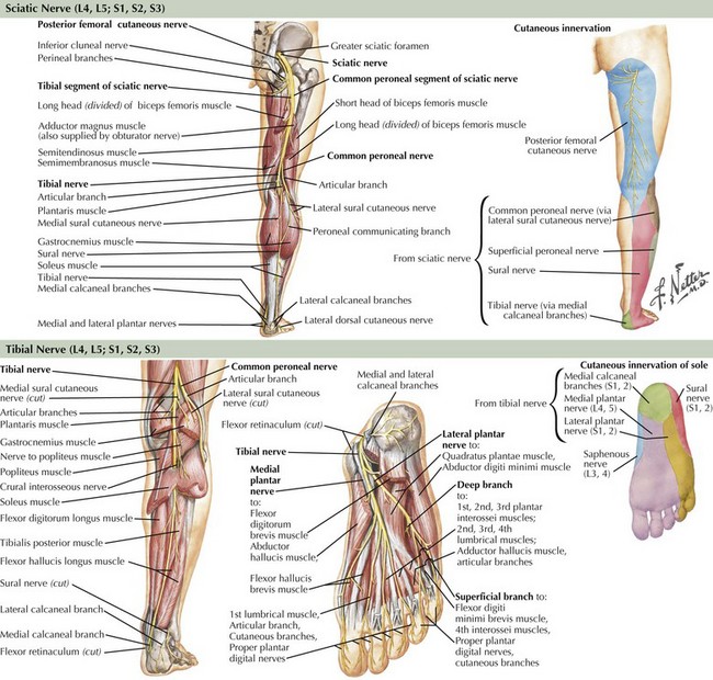
The sciatic nerve is the body’s largest nerve, receiving contributions primarily from the L5, S1, and S2 nerve roots, but also carrying L4 and S3 fibers (Fig. 66-1). It has two primary divisions: the laterally situated more superficial peroneal nerve and the more medially placed tibial nerve (see Fig. 66-1). These separate into two distinct nerves in the mid- to distal thigh. The sciatic nerve and its branches innervate the hamstrings (biceps femoris, semimembranosus, and semitendinosus muscles), distal adductor magnus, anterior and posterior lower leg compartments, and intrinsic foot musculature. Through sensory branches of the tibial nerve (sural, medial and lateral plantar, and calcaneal) and the superficial peroneal nerve, the sciatic nerve also supplies sensation to the skin of the entire foot and the lateral and posterior lower leg.
Peroneal Neuropathies
Etiology
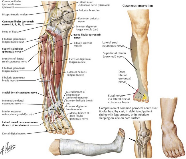
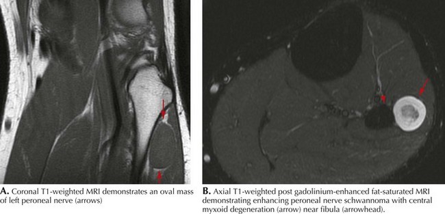
Common peroneal neuropathy is the most frequent lower extremity mononeuropathy. The common peroneal nerve is most susceptible to external compression at the fibular head, where it is very superficial (Fig. 66-2). Predisposing causes include recent substantial weight loss, habitual leg crossing, or prolonged squatting. External devices such as casts, braces, and tight bandages can also cause peroneal neuropathy. Diabetes mellitus, vasculitis, and rarely hereditary tendency to pressure palsy (HNPP) are other etiologic conditions. An acute anterior or lateral compartment syndrome below the knee can also lead to acute common, deep, or superficial peroneal neuropathies. Patients with insidious onset and progressive course require evaluation for mass lesions, including a Baker cyst or ganglion, osteoma, or schwannoma (Fig. 66-3). The common peroneal nerve is sometimes injured iatrogenically. Knee positioning and padding to decrease pressure on the peroneal nerve in the operating room and intensive care unit are important to prevent an acute compression neuropathy. Rarely, laceration of the peroneal nerve occurs with arthroscopic knee repair or direct penetrating trauma.
Clinical Presentation
Most peroneal neuropathies involve the common peroneal nerve at the fibular head causing weakness of foot dorsiflexion and eversion (see Fig. 66-1). Ambulation reveals a “steppage gait” with compensatory hip and knee flexion in order to lift the foot off the floor. The foot might hit the floor with a slap, as the patient has poor control over its movements. With the less frequently occurring deep peroneal neuropathies, there is weakness of the tibialis anterior, extensor hallucis, extensor digitorum longus, and extensor digitorum brevis. Primary superficial peroneal neuropathies cause weakness of the peroneus longus and brevis muscles, which are mainly responsible for foot eversion.


