Fig. 55.1
Rhinitis medicamentosa. Axial (a) and coronal (b) CT images show marked mucosal thickening of the bilateral inferior turbinates (arrows), with resultant nasal airway obstruction
55.4 Differential Diagnosis
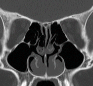
Allergic and infectious rhinitis can also lead to swelling of the nasal mucosa and secretions within the nasal cavity and can have similar appearances on CT (Fig. 55.2). The distinction between allergic or infectious rhinitis and rhinitis medicamentosa depends on the clinical scenario. Unilateral enlargement of the nasal turbinate mucosa can occur naturally during the process of the nasal cycle (Fig. 55.3), in which there is alternating turgescence of the mucosa on one side versus the other. Although the middle turbinate can appear enlarged with concha bullosa, it is a developmental variant that affects the bony anatomy rather than the mucosa. The pneumatized middle turbinates are readily identified on CT (Fig. 55.4). Other potential mimics of turbinate hypertrophy are various nasal cavity neoplasms, including recurrent tumors that occupy the space of resected turbinates (Fig. 55.5).


Fig. 55.2


Allergic hypertrophic rhinitis. Coronal CT image shows swelling of the bilateral inferior turbinate mucosa. There are also bilateral concha bullosa, which are partially opacified
Stay updated, free articles. Join our Telegram channel

Full access? Get Clinical Tree


