14 Nerve roots
Development of the Spinal Cord
Cellular differentiation
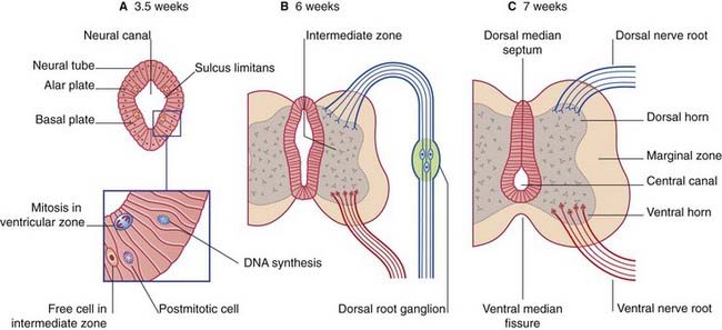
The neural tube of the embryo consists of a pseudostratified epithelium surrounding the neural canal (Figure 14.1A). Dorsal to the sulcus limitans, the epithelium forms the alar plate; ventral to the sulcus it forms the basal plate.
The microglial cells of the CNS are derived from basophil cells of the blood.
Enlargement of the intermediate zone of the alar plate creates the dorsal horn of gray matter. The dorsal horn receives central processes of dorsal root ganglion cells (Figure 14.1B). As explained in Chapter 1, the ganglion cells derive from the neural crest.
Partial occlusion of the neural canal by the developing dorsal gray horn gives rise to the dorsal median septum and to the definitive central canal of the cord (Figure 14.1C).
Enlargement of the intermediate zone of the basal plate creates the ventral gray horn and the ventral median fissure (Figure 14.1C). Axons emerge from the ventral horn and form the ventral nerve roots.
In the outermost, marginal zone of the cord, axons run to and from spinal cord and brain.
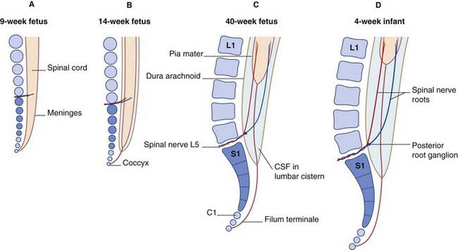
Ascent of the spinal cord (Figure 14.2)
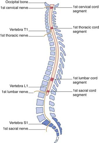
In consequence of greater ascent of the lower part of the cord compared to the upper part, the spinal nerve roots show an increasing disparity between their segmental levels of attachment to the cord and the corresponding vertebral levels (Figure 14.3).
Neural arches
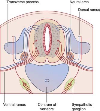
During the fifth week, the mesenchymal vertebrae surrounding the notochord give rise to neural arches for protection of the spinal cord (Figure 14.4). The arches are initially bifid (split). Later, they fuse in the midline and form the vertebral spines.
Conditions where the two halves of the neural arches have failed to unite are collectively known as spina bifida (Clinical Panel 14.1).
Clinical Panel 14.1 Spina bifida
Among the more common congenital malformations of the CNS are several conditions included under the general heading spina bifida. The ‘bifid’ effect is produced by failure of union of the two halves of the neural arches, usually in the lumbosacral region (Figure CP 14.1.1)
In spina bifida cystica, a meningeal cyst protrudes through the vertebral defect. In 10% of these cases, the cyst is a meningocele containing no nervous elements (B). In 90%, unfortunately, the cyst is a meningomyelocele, containing either spinal cord or cauda equina (C); the lower limbs, bladder, and rectum are paralyzed, as in the case illustrated in Figure CP 14.1.2, and meningitis is likely to supervene sooner or later. To make matters worse, an Arnold–Chiari malformation (Ch. 4) is almost always present as well.

Stay updated, free articles. Join our Telegram channel

Full access? Get Clinical Tree


