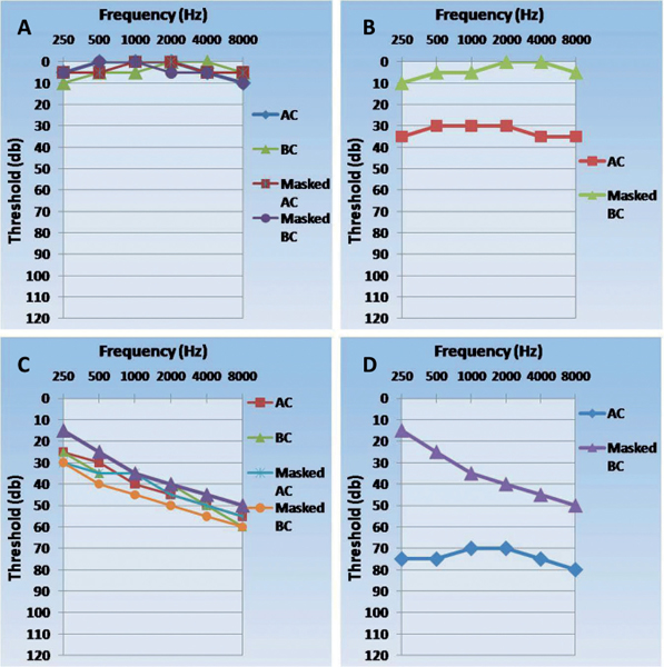
22 What are the two sensory divisions of cranial nerve VIII? Cochlear and vestibular divisions1 Within what bony structure does the membranous labyrinth lie? The bony labyrinth, contained within the petrous portion of the temporal bone2 Describe the structures traversed by a sound wave on its way to the cochlea. The wave first traverses the pinna and enters the external auditory canal to strike the tympanic membrane. Vibrations of the tympanic membrane are transmitted by three middle ear ossicles (malleus, incus, stapes) to the oval window, where the sound is then transmitted into the inner ear and cochlea. Within the cochlea, the sound is transmitted through the perilymph within the scala vestibuli and propagates along the cochlea, which contains hair cells that transduce the mechanical vibrations caused by sound into an electrical signal. What middle ear structure seals the oval window? The stapes What organ within the cochlea allows for the transduction of a mechanical sound signal into an electrical impulse? The organ of Corti. The tectorial membrane overlying the cochlear hair cells vibrates against the kinocilia of each hair cell, mechanically distorting the hair cells and resulting in depolarizations. What causes mechanical deflection of the hair bundles in the organ of Corti? Differential fluid pressure between the scala vestibula and scala tympani Does the kinocilium directly participate in mechanoelectrical transduction? No. The kinocilium transmits forces to the stereocilia in a hair bundle; they participate in transduction. Kinocilium often possess a core composed of microtubules, unlike stereocilium, which are composed of actin microfilaments. The more rigid composition of the former allows it to act as a moment arm. Describe the afferent and efferent innervations pattern in the organ of Corti. Afferent axons of cochlear ganglion cells traveling to the cochlear nucleus originate mainly with inner hair cells, whereas efferent axons from the superior olivary complex terminate mostly in the outer hair cells. How is the sensitivity of the auditory system modified? The tympanic membrane can be drawn taut under the influence of the trigeminal nerve (CN V), making it less sensitive to sound. The movement of the stapes in response to sound may be decreased by the stapedius muscle under the influence of the facial nerve (CN VII). The cochlea is tonotopically organized (as are all auditory connections between the cochlea and cortex). In what portion of the cochlea do high-frequency sounds reach their peak? Low-frequency sounds? High-frequency sounds reach their peak at the base of the cochlea (near the oval window). Low-frequency sounds reach their peak at the cochlear apex (near the round window). Where do the cell bodies of the sensory neurons innervating the organ of Corti lie? In the cochlear ganglion Where do the axons of the cochlear ganglion project? In the cochlear division of CN VIII to reach the ventral and dorsal cochlear nuclei near the pontomedullary junction of the brainstem Describe the course of the second-order axons in the auditory pathway. The ventral and dorsal nuclei project axons that travel rostrally on both sides of the brainstem (crossed and uncrossed fibers) through the trapezoid body (in the caudal pons) and within the lateral lemnisci (midpons) to the inferior colliculi (midbrain), where they project to the medial geniculate bodies bilaterally. What is the role of the medial geniculate bodies? Where do they project? The medial geniculate bodies (MGBs) serve as the thalamic auditory relay. Third-order neurons in each MGB receive auditory information from both ears and transmits this information via the sublenticular portion of the internal capsule to the auditory cortex in the posterior portion of the superior temporal gyrus (Brodmann’s area 41). What audiological deficit would you expect with a lesion to the lateral lemniscus, CN VIII, or more distally in the auditory system? Contralateral sensorineural deafness (CN VIII lesions would also likely cause vertigo, balance difficulties, and nystagmus due to damage to the vestibular portion). What neurological deficit would you expect with a lesion to the MGB? Each MGB receives input from both ears, thus both ears would still retain some auditory function bilaterally. A lesion to what structure can result in auditory hallucinations? Pons What are the saccule and utricle? The portions of the membranous labyrinth governing sensation of horizontal and vertical acceleration. They contain hair cells and otoliths (calcium carbonate) clustered in macular areas. What are the ampullae? Enlargements of the three semicircular canals. Each contains a crista ampullaris and together the three are sensitive to rotation on any axis. A gelatinous cupula covers each ampulla and mechanically distorts hair cells, producing depolarizations in response to rotation. Where do the cell bodies of the primary sensory neurons for the vestibular system lie? Where do they project? In the vestibular ganglion. As with the cochlear ganglion, these cells are bipolar, sending peripheral branches to the saccule, utricle, and ampullae, and central branches that travel with the auditory branches of the cochlear ganglion in CN VIII to synapse in the vestibular nuclei in the caudal pons. Where do fibers of the vestibular nuclei project? 1. Flocculonodular cortex of the cerebellum 2. Ipsilateral spinal cord (vestibulospinal tract) 3. To motor nuclei of extraocular muscles (vestibulo-ocular reflex) and spinal cord ventral horn via the medial longitudinal fasciculus 4. To parietal cortex via the ventral posterolateral nucleus (VPL) of the thalamus Which cranial nerves are involved in the vestibulo-ocular pathway? III, VI, and VIII What is nystagmus? A rhythmic movement of the eyes involving a fast, jerking motion in one direction and a slow, pulling motion in the other; can be induced physiologically or may be a sign of a lesion in the vestibular pathway3 Cool water placed within the external ear canal causes a nystagmus (the fast-component) to which side? Warm water? Remember the mnemonic COWS (with regard to fast-component): Cool water produces a nystagmus to the Opposite side Warm water produces a nystagmus to the Same side4 CNS nystagmus may often be differentiated from peripheral vestibular nystagmus by the presence or absence of what symptom? Vertigo is often present with peripheral vestibular nystagmus, but not with central nystagmus. What is audiometry? The study of a subject’s ability to detect audible noises of different frequencies (Hz) and intensities (decibels). Both air conduction and bone conduction may be measured (allowing for the discrimination of the etiology of hearing loss). What is “threshold”? The softest intensity level at which a particular single frequency sound can be detected by a subject 50% of the time. What is an audiogram? A graphic display of a subject’s threshold values as a function of the different frequencies tested.5 Between which frequencies does normal human conversation occur? 250 to 8000 Hz What is the range of frequencies detectable to most humans? 20 to 20,000 Hz Masking procedures are techniques used in attempts to prevent the nontested ear from responding to signals presented to the test ear.2 What are some causes of conductive hearing loss? Cerumen or foreign-body buildup in external auditory canal(s), otitis externa or media, and otosclerosis What is the most common cause of sensorineural deafness? Loss of cochlear hair cells. May also be due to CN VIII damage (e.g., acoustic neuroma) or damage to central auditory pathways. Fig. 22.1 (A) Normal audiogram. (B) Conductive hearing loss audiogram. (C) Sensorineural hearing loss audiogram. (D) Mixed hearing loss audiogram.
Neuro-Otology
22.1 Basic Concepts
22.2 Audiogram and Audiology Examination
Neuro-Otology
Only gold members can continue reading. Log In or Register to continue


Full access? Get Clinical Tree


