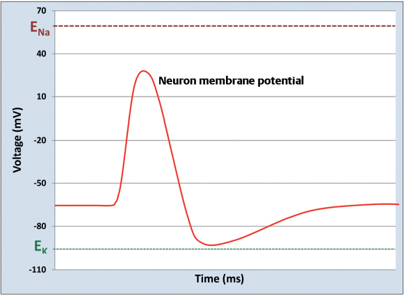
18 What are the two major types of cell death?1 Necrosis: cell death resulting from injury (ischemia, infection, toxins, immune reactions, hypoglycemia, etc.) Apoptosis: programmed cell death, occurring through an internally controlled suicide mechanism Give examples of adaptations a cell may undergo in response to injurious stimuli. Hyperplasia, hypertrophy, atrophy, and metaplasia Describe the neuronal cell response to injury. 1. Cell swelling: nucleus, nucleolus, and cell body swell as an initial response to insult 2. Chromatolysis: Nissl substance (ribosomal RNA) dissolves, allowing protein production to take place. Mitochondria begin to swell. Smooth endoplasmic reticulum (ER) proliferates to produce new cell membrane and myelin. 3. Recovery or death What excitotoxins are generated in addition to free radicals and cytotoxic cytokines during necrotic neuronal cell death following acute ischemia or traumatic injury? What type of cell death occurs in the penumbra after a focal cerebral ischemia? Apoptosis What are the histological features of apoptosis? During apoptosis, the cytoplasm condenses, mitochondria and ribosomes aggregate, the nucleus condenses, and chromatin aggregates.1 What are the histological features of necrosis? Mitochondrial and nuclear swelling, dissolution of organelles, and condensation of chromatin around the nucleus In chronic neurodegenerative diseases such as Huntington’s, amyotrophic lateral sclerosis, and Alzheimer’s disease, what is the predominant form of cell death? Apoptosis4 Removal of which biochemical factor from neuronal culture media has been shown to promote apoptosis? Neurotrophic factors (brain-derived neurotrophic factor and neurotrophin-3) How long after an axon in the PNS is sufficiently injured does degeneration begin? Within hours the axon and surrounding myelin begin to fragment and ultrastructural evidence of neurotubule and neurofilament disarray can be seen. By 48 to 96 hours postinjury, axonal continuity has been reduced such that impulses cannot be conducted.5 At what rate do regenerating axons grow? Approximately 1 mm/day5 Why do axons fail to regenerate and reinnervate their target organ or tissue in the CNS? The presence of environmental inhibitors (e.g., myeline-associated inhibitors and chondroitin-associated proteoglycans) as well as the failure of CNS neurons to upregulate growth-associated genes limits axonal regeneration in the CNS.6 What are the two principal types of cells in the nervous system? Neurons and glial cells Describe the structure of a neuron. 1. The cell body, or soma, maintains the metabolic needs of the cell and houses the nucleus, Golgi apparatus, Nissl substance (ribosomal RNA), cytoskeleton, and mitochondria. 2. Dendrites are tapered extensions of the cell body designed to collect information from other neurons and the environment. 3. Axons are extensions that conduct and convey information to other neurons by excreting neurotransmitters from vesicles contained within the axon into a synapse, where the neurotransmitters travel to another neuron and transmit a particular signal. True or false: As in all other cells, insulin is required for glucose uptake into neurons. False. Insulin is required for efficient glucose uptake into the majority of all bodily cells (primarily muscle and adipose tissue) but is not required for uptake into neurons. Which type of neuron is the most numerous in the nervous system: sensory neurons, motor neurons, or interneurons? Interneurons What filamentous structures are responsible for the shape of a neuron? Microtubules, neurofilaments, and actin microfilaments create the cytoskeleton of a neuron. Microtubules consist of 13 protofilaments in a 25-nm tubular array with each protofilament consisting of several pairs of α and β tubulin. Neurofilaments (similar to intermediate filaments outside the nervous system) are 10-nm polymers of cytokeratins. Microfilaments are 7-nm pairs of polymerized actin. Which filamentous protein polymers serve as the substrate for organelle transport within neurons? Microtubules Which filamentous protein polymers are important for movement of the advancing tip of growing axons? Microfilaments (actin) Where are axons located in the nervous system? Cell bodies? Axons are located in the central nervous system (CNS) white matter and in peripheral nerves in the peripheral nervous system (PNS). Cell bodies are housed in CNS gray matter and PNS ganglia. How fast is slow axonal transport? Fast axonal transport? Slow axonal transport occurs only in the anterograde direction and consists of a slower component (0.2 to 2.5 mm/day) and a faster component (0.4 to 5 mm/day). Fast axonal transport occurs in both anterograde and retrograde directions at greater than 400 mm/day.7 Which microtubule-associated ATPase is responsible for anterograde transport? Kinesin7 Can anterograde transport occur in nerves severed from their respective cell bodies? Yes. Anterograde transport does not rely on the presence of a cell body. This is the basis for in vitro motility assays. Which microtubule-associated ATPase is responsible for retrograde transport? MAP-1C, a microtubule-associated ATPase7 similar to dyneins in cilia and flagella What is the typical resting membrane potential of a neuron? −60 mV to −70 mV7 What is an ion’s equilibrium potential? The membrane potential resulting from a membrane selectively permeable to the given ion if allowed to reach a steady state (or equilibrium) in which both concentration gradients and charge forces counterbalance one another What is the primary determinant of cell membrane potential? The permeability of the cell membrane to different ions. The cell membrane potential will move toward the equilibrium potential for ions to which it is relatively more permeable. This resting potential is due to the relatively high permeability of the resting neuronal cell membrane to what ion? K+, which has an equilibrium potential of −94 mV. There is also a relatively small permeability of the resting neuronal cell membrane to Na+. An equation describing the relationship between the predicted membrane potential and variables such as the valence of an ion and the concentrations of the ion inside and outside the cell. For a membrane permeable only to K+ at a temperature of 37°C: Vm = 62log10 [K+]outside cell/[K+]inside cell (Eq. 18.1) What are the three states assumed by voltage-gated sodium channels? They become activated in response to depolarization, resulting in an influx of Na+ and further depolarizing the neuron. After being activated, they quickly become inactivated and unresponsive to further polarization (refractory period). The neuron is subsequently repolarized by the opening of K+ channels, which restores the voltage-gated sodium channels back to their resting state.8 Fig. 18.1 Illustration of a typical neuron action potential in relation to the equilibrium potentials of sodium and potassium.
Neurobiology
18.1 Neuronal Death and Regeneration
18.2 Neuron/Axon Structure
18.3 Action Potential
Neurobiology
Only gold members can continue reading. Log In or Register to continue


Full access? Get Clinical Tree


