Neurologic Examination: Grading Scales


Neurologic Examination: Grading Scales


Charles H. Tator


The clinical neurologic examination and imaging of the spine and spinal cord are the most important components of the assessment of patients with acute cervical spinal cord injury (SCI). Accurate management in the acute and subsequent stages including rehabilitation depends on the findings of the neurologic examination. The neurologic examination has several components, which must be performed in a consistent and reproducible fashion, so that each phase of care can be compared with the previous, in order to assess any positive or negative trends in response to management. The neurologic examination of cervical SCI is similar to the examination performed for other levels of spinal cord or cauda equina injury in that it includes motor, sensory, reflexes, and sphincter function. However, there are unique aspects of the neurologic assessment of cervical SCI such as the zone of partial preservation, the potential involvement of respiratory function, and the propensity for major autonomic dysfunction.

The neurologic findings allow the examiner to define the neurologic level of injury and to classify and grade the extent and severity of the neurologic injury according to one of several recognized systems of assessment, which are based solely on the assessment of neurologic function. To date, only systems of scoring based on clinical neurologic function have been proven to be of value (1), although in the future it is hoped that imaging or electrophysiologic features will be sufficiently rigorously defined to allow further classification of SCI based on these modalities. Recent studies show promise in categorizing the severity of cervical compression SCI based on T1 and T2 midsaggital magnetic resonance imaging (2,3). Although there is promise that electrophysiologic assessment will become useful for assessment of the severity of acute SCI, its current role is for monitoring the subacute and chronic phases of injury (4,5), although it is of value for intraoperative monitoring in SCI (6).

An accurate system of clinical neurologic assessment permits the development of a rational treatment plan, allows reliable and consistent serial monitoring of the patient during the acute and rehabilitative phases of care, and provides useful data for the early determination of prognosis. Furthermore, the system of neurologic assessment and classification must be sufficiently reliable to allow accurate comparison of serial observations by the same or different observers. Many systems of classifying and scoring the extent and severity of the clinical neurologic deficit have been developed, and this chapter provides a review and critical analysis of the various systems, both simple and complex, that have been used. Since 1992, the system used in most countries is the International Standards for Neurological and Functional Classification of Spinal Cord Injury (ISCSCI), initially developed in the 1980s by the American Spinal Injury Association (ASIA). Then, in the early 1990s, ASIA convened consensus meetings of representatives from several disciplines and from several countries involved in the management of patients with acute SCI, and in 1992, ASIA along with the International Medical Society of Paraplegia, now renamed the International Spinal Cord Society (ISCoS), published the ISCSCI (7), with the most recent update in 2006. This system perhaps should be used by all physicians, surgeons, and other health care professionals managing patients with acute SCI.


SYSTEMS OF NEUROLOGIC CLASSIFICATION AND SCORING

The systems of neurologic classification can be divided into simple and complex systems. Simple systems primarily describe whether the neurologic injury is complete or incomplete and indicate the anatomical location in the cord with the most prominent neurologic injury (Table 14.1). In contrast, complex systems provide a detailed account of the neurologic injury in terms of a standardized grading of motor and sensory function (Table 14.2).


SIMPLE SYSTEMS FOR NEUROLOGIC CLASSIFICATION AND SCORING


COMPLETE VERSUS INCOMPLETE SPINAL CORD INJURY SYNDROMES

One of the simplest and most useful systems is the grading of patients into complete and incomplete neurologic injuries. Historically, this was probably the first system used
and antedates all of the more complex systems. A complete injury had the following: complete loss of voluntary motor function below the level of the injury, complete absence of somatic sensation below the level of the injury, and complete loss of voluntary control of bowel and bladder function. Later, other features were added to the definition of the complete syndrome such as the presence of abnormal reflexes including priapism and the early return of the bulbocavernosus reflex. In contrast, a patient was neurologically incomplete when there was any residual voluntary motor or sphincter function or sensation below the level of the injury. As shown in Table 14.1, a large number of incomplete syndromes have been described, generally based on the anatomical location of the maximal cord injury in the transverse plane.


TABLE 14.1 Simple Systems for Neurologic Classification of Acute Cervical SCI: Complete and Incomplete Neurologic Syndromes


1. Complete SCI Syndromes—ASIA Grade A


(a) Unilevel: no zone of partial preservation


(b) Multilevel: with zone of partial preservation


2. Incomplete SCI Syndromes—ASIA Grades B, C, or D


(a) Cervicomedullary syndrome


(b) Central cord syndrome


(c) Anterior cord syndrome


(d) Posterior cord syndrome


(e) Brown-Séquard syndrome


3. SCIWORA—ASIA Grades A, B, C, or D. Mainly children and youth


4. SCIWORET ASIA Grades A, B, C, or D. Mainly adults


5. Reversible or Transient Syndromes


(a) Cord concussion


(b) Burning hands syndrome


(c) Contusion cervicalis


(d) Hysteria


(e) Malingering


6. Spinal Cord Trauma Syndromes Without Direct Cord Injury


The major advantages of these simple systems are simplicity and usefulness for estimating prognosis in the acute phase. Although prognostic accuracy for complete injuries is reasonably good, prognostic accuracy for incomplete injuries is poor: Complete injuries have virtually no likelihood of major neurologic recovery (such as recovery of ambulation), while incomplete injuries can recover almost to normal. For example, Hansebout (8) showed that only a very small number of complete injuries recover ambulation. He analyzed several large series of acute SCI patients and found that a small percentage of initially complete cases (usually 1% to 2%) had significant recovery of distal cord function. However, it can be argued that even these rare cases of recovered complete injuries were misdiagnoses due to factors discussed in detail below such as inebriation, sedative or other drug effects, spinal shock, uncooperativeness, or a concomitant head injury. More recent estimates of the number of recovered complete cord injuries are similar to Hansebout’s, and these include the large National Acute Spinal Cord Injury Study (NASCIS) (9) and Sygen (10) pharmacotherapy trials and the large database of the US model systems (11). They show that some complete patients can recover significant distal cord function, even in the absence of all of the factors listed above that can interfere with precise, early classification. In contrast, neurologically incomplete injuries have a much better prognosis for recovery and a very large range of recovery from minimal to almost total recovery (12), and thus, prognostic accuracy in individual cases in the early phase is very poor (13,14).


TABLE 14.2 Complex Systems for Neurologic Classification and Scoring in Acute Cervical SCI


System


Features


Advantages


Disadvantages


Frankel


5 Severity grades


Simple, easy to understand


Cannot quantify recovery Imprecise definitions of grades A, C, and D Ceiling effect


Sunnybrook


10 Severity grades 17 Neurologic changes


More accurate Initial grading Allows quantification of recovery in individuals and groups


No numerical scores of motor and sensory function


ASIA


5 Severity grades


Improved Definition of Complete Spinal Cord Injury Allows quantification of neurologic scores


Ceiling effect


Benzel


7 Severity grades Sphincter control


Cannot be used for initial examination


In addition to prognostic imprecision, there are other significant disadvantages to the simple systems including lack of precision of what constitutes a complete injury. For example, until recently, there was no commonly accepted definition of a complete injury, and some features meant to refine the definition of completeness such as the return of the bulbocavernosus reflex are unreliable. Furthermore, the simple systems do not quantify the severity of the neurologic deficit in an individual patient or groups of patients and are of minimal value for serial observation in a given patient. Therefore, the simple systems cannot be used alone and must be supplemented by one of the complex methods described below.


INCOMPLETE SYNDROMES OF ACUTE CERVICAL SPINAL CORD INJURY

Table 14.1 shows the large number of incomplete neurologic syndromes in cervical SCI named according to the
presumed location of the maximal injury in the transverse plane of the spinal cord (15) (Figs. 14.1, 14.2, 14.3, 14.4 and 14.5). It is useful clinically to categorize incomplete patients on the basis of the anatomical location of the cord injury because it provides information about the mechanism of injury and influences selection of treatment. These incomplete syndromes have differing prognoses for recovery, and therefore, categorization provides prognostic information.


Cervicomedullary Syndrome

A high proportion of injuries to the upper cervical cord are associated with damage to the medulla, hence the term cervicomedullary syndrome (16,17) or cruciate paralysis is used (18). These injuries may extend from the pons to C4 or even lower in the cord and are due to either direct injury, stretching, or vertebral artery injury. The essential clinical features are respiratory insufficiency or arrest, hypotension, varying degrees of tetraparesis, sensory changes from C1 to C4, and sensory loss over the face in the onion skin or Dejerine pattern. There is often greater arm than leg weakness. The more rostral the lesion, the more severe the manifestations such as in patients with atlanto-occipital dislocation. The mechanisms of cord injury include traction injury, as in severe atlantoaxial dislocation, and anteriorposterior compression from burst fracture, odontoid fracture, or ruptured disk. Skilled, prompt first aid and triage have increased the likelihood of survival, and thus, more of these cases live to reach hospital than previously.


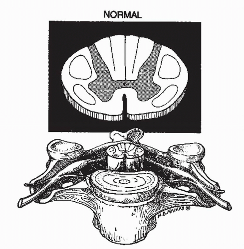
Figure 14.1. The normal spinal cord and spinal column. The normal relationships between the spinal cord, spinal column, and nerve roots are depicted in the mid cervical region. For clarity the durra has been omitted. In the upper diagram, the gray matter is finely stippled, and the corticospinal and spinothalamic tracts are outlined. The intervertebral disk is shown. Reprinted with permission from Narayan et al. Neurotrauma. New York: McGraw-Hill, 1996, Chapter 75.


Acute Central Cord Syndrome

Schneider described the acute central cervical cord syndrome characterized by a disproportionately greater loss of motor power in the upper than lower extremities and variable sensory loss. He hypothesized that acute compression was an etiologic factor in many cases (19) (Fig. 14.2). There are many similarities between the central cord syndrome and the syndrome of cruciate paralysis, and it may be very difficult to differentiate the two clinically (20). There is recent evidence that these syndromes characterized by greater arm than leg weakness are not due to the presumed differing locations of the arm and leg fibers in the corticospinal tract as previously thought (21). The evidence from careful correlation of the clinical findings, MR imaging, and pathology obtained at autopsy (22) and mounting evidence that the lateral corticospinal tract subserves mainly distal limb musculature account for greater hand dysfunction from major damage to this tract. The incidence of the central cord syndrome varies from 1% to 4% of SCI and is the most common of these identifiable syndromes (23). The prognosis after central cord injury varies considerably, but many patients remain with permanent weakness, spasticity, and proprioceptive loss, especially in the hands (24). There is recent evidence that MRI T2 signal change may be an important diagnostic predictor (25).


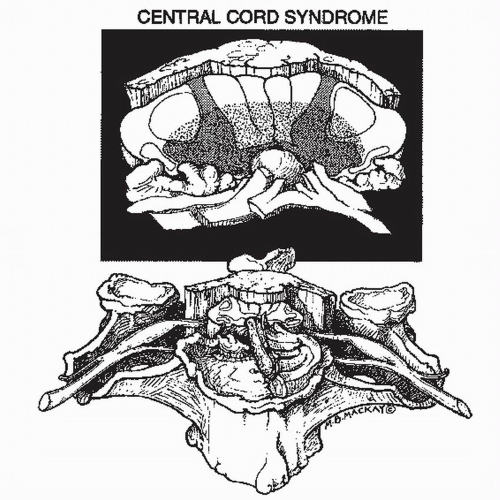
Figure 14.2. Central cord syndrome. The drawing depicts a case of cervical spondylosis with osteoarthritis of the cervical spine including anterior and posterior osteophytes and hypertrophy of the ligamentum flavum. Superimposed is an acute hyperextension injury, which has caused rupture of the intervertebral disk and infolding of the ligamentum flavum. The spinal cord is compressed anteriorly and posteriorly. The central portion of the cord shown in rough stippling sustained the greatest damage. The damaged area includes the medial segments of the corticospinal tracts presumed to subserve arm function. Reprinted with permission from Narayan et al. Neurotrauma. New York: McGraw-Hill, 1996, Chapter 75.


Anterior Cord Syndrome

This syndrome was also originally described in the setting of acute cervical trauma by Schneider and Thompson (26) who presented two cases of “immediate complete paralysis with hyperesthesia at the level of the lesion and an associated sparing of touch and some vibration sense.” In Schneider’s view, this was “a syndrome for which early operative intervention is indicated.” The anterior aspect of the cord is damaged, and in severe cases, there may only be sparing of the posterior columns (Fig. 14.3). Less severe cases may have partial retention of motor function due to sparing of some fibers in the lateral corticospinal tracts.


Posterior Cord Syndrome

This is the least common type of incomplete syndrome (23). Many observers including the author have doubted its existence. It is postulated that it occurs after major destruction of the posterior aspect of the cord but with some residual functioning spinal cord tissue anteriorly (Fig. 14.4). Thus, clinically, the patient would have retained spinothalamic function but would have lost movement and proprioception due to damage to the posterior half of the cord including the corticospinal tracts and posterior columns.


Brown-Séquard Syndrome

The Brown-Séquard syndrome is caused by a lesion of the lateral half of the spinal cord (Fig. 14.5) and is characterized by ipsilateral motor and proprioceptive loss and contralateral pain and temperature loss. The syndrome can be associated with a variety of mechanisms of injury, most frequently hyperextension, although also with flexion, locked facets, and compression fractures.


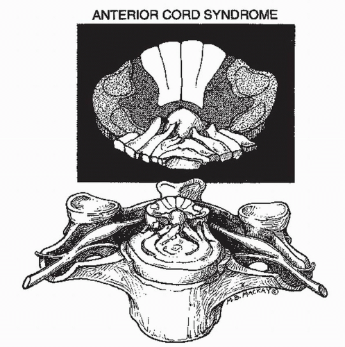
Figure 14.3. Anterior cord syndrome. A large disk herniation is shown compressing the anterior aspect of the cord and resulting in damage (rough stippling) to the anterior and lateral white matter tracts and to the gray matter. The posterior columns remain intact. Reprinted with permission from Narayan et al. Neurotrauma. New York: McGraw-Hill, 1996, Chapter 75.

The Brown-Séquard syndrome may be present in the acute phase or may become apparent several days after injury as a gradual evolution from a bilateral incomplete injury. Hybrid combinations of Brown-Séquard and other incomplete syndromes may occur (23). For example, the author has seen frequent examples of central cord injuries that are quite asymmetric with the more severely damaged side of the cord showing features of a Brown-Séquard syndrome. Patients with this syndrome have better prognosis for ambulation than many of the other syndromes (23).


SPINAL CORD INJURY WITHOUT RADIOLOGIC ABNORMALITY AND SPINAL CORD INJURY WITHOUT RADIOLOGIC EVIDENCE OF TRAUMA

The syndrome of SCI without radiologic abnormality (SCIWORA) is more common than in adults and represents a significant percentage of pediatric SCI (27). Children are more susceptible to this injury presumably because of the laxity of their spinal ligaments and the weakness of their paraspinal muscles. Children with SCIWORA tend to have less severe SCI than those with definite evidence of bony injury, although complete injuries have been described. By
definition, the negative radiologic examination includes only plane films and tomography, either conventional or CT. If a negative MR was included in the definition, the number of cases would diminish dramatically because of the extreme sensitivity of MR for detecting even mild cord injury such as T2 signal change and spinal column injury such as torn ligaments and ruptured disks. True SCIWORA can also occur in adults but is much less frequent than the syndrome of acute SCI without radiologic evidence of trauma (SCIWORET) (28). Cervical spondylosis is the most commonly associated condition in adults with SCIWORET, but other conditions including spinal stenosis, ankylosing spondylitis, disk herniation, and nucleus pulposus embolism may on rare occasion be associated with SCI and not show radiologic evidence of trauma (28). Thus, in SCIWORET, there are often abnormal radiologic exams, but no evidence of trauma. Prior to the use of CT or MRI in spinal trauma, the incidence of SCIWORET in SCI in adults was approximately 14% (15). The addition of CT has reduced the incidence of SCIWORET to about 5%. One study showed that many patients with SCIWORET have demonstrable compression by myelography or MRI and should be considered for early surgical decompression (29). It is likely that very few SCIs will remain undetected by MR because of its high sensitivity for detecting mild SCI and nonbony spinal column lesions.


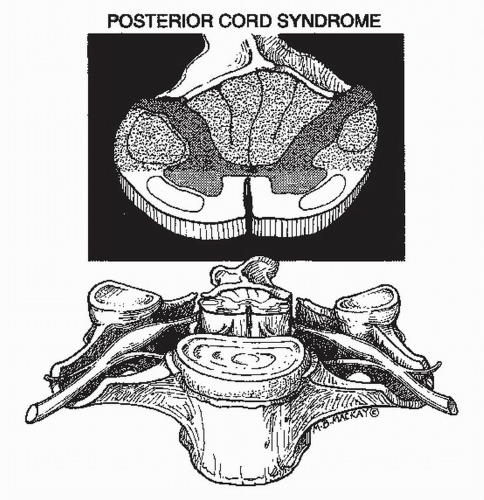
Figure 14.4. Posterior cord syndrome. A laminar fracture is depicted with anterior displacement of the fractured bone and compression of the posterior aspect of the spinal cord. The damaged area of the cord (roughly stippled in the upper diagram) includes the posterior columns and the posterior half of the lateral columns including the corticospinal tracts. Reprinted with permission from Narayan et al. Neurotrauma. New York: McGraw-Hill, 1996, Chapter 75.


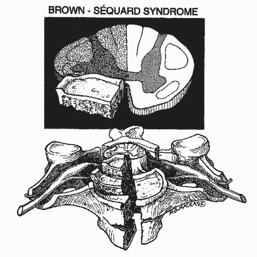
Figure 14.5. Brown-Séquard syndrome. A burst fracture is depicted with posterior displacement of bone fragments and disk, resulting in unilateral compression and damage (rough stippling) to one-half of the spinal cord. Reprinted with permission from Narayan et al. Neurotrauma. New York: McGraw-Hill, 1996, Chapter 75.

Only gold members can continue reading. Log In or Register to continue

Stay updated, free articles. Join our Telegram channel

Jun 29, 2016 | Posted by in NEUROLOGY | Comments Off on Neurologic Examination: Grading Scales

Full access? Get Clinical Tree

Get Clinical Tree app for offline access