Neuromuscular Diseases


Neuromuscular Diseases


Dianna Quan

Steven P. Ringel


Disorders that result from abnormalities of spinal cord motor neurons, peripheral nerve, neuromuscular junction, or muscle constitute “neuromuscular disease.” Weakness is a common symptom, but unlike central nervous system (CNS) disorders, neuromuscular diseases may be accompanied by muscle atrophy and diminished muscle tone. Patients also may develop muscle pain, stiffness, cramps, twitching, limb deformities, or myoglobinuria.

This chapter begins with an overview of the variable clinical presentations of neuromuscular disease and a description of the tests commonly used to aid in the diagnosis of neuromuscular conditions. Subsequent sections include more detailed descriptions of specific neuromuscular disorders, excluding disorders of peripheral nerve, which will be covered in a separate chapter. For each condition, pertinent diagnostic investigations including genetic testing (Table 17.1), outpatient treatment principles (Table 17.2), special problems in managing hospitalized patients, and suggestions on when to refer to a neurologist will be discussed.


CLINICAL PRESENTATIONS


Weakness

▪ SPECIAL CLINICAL POINT: A useful clinical generalization for neuromuscular disorders is as follows:all motor neuron, neuromuscular junction, and muscle diseases have no sensory changes accompanying weakness.

Coexisting sensory loss, dysesthesias, or paresthesias strongly suggest a peripheral nerve disorder (see Chapter 18, Peripheral Neuropathy).


TABLE 17.1 Gene Abnormalities in Neuromuscular Disease


Disorder


Chromosome


Recessive (R) or Dominant (D)


Gene Product


Special Features


Motor Neuron Disease


Spinal muscular atrophy


SMA 1,2


5


(R)


SMN and rarely NAIP protein in SMA 1,2,3


SMN protein mutation in 95% of Type 1,2,3


SMA 3


5


(R) and (D)


SMA 4 (adult)


Unknown


(R) and (D)


SMN defect in some


Clinical overlap with progressive muscular atrophy


Spinal and bulbar muscular atrophy (Kennedy’s disease)


X


(R)


Androgen receptor


Androgen receptor gene enlarged (multiple CAG repeats)


Amyotrophic lateral sclerosis


Sporadic (90%-95%)


No known defect


None known


See text


Familial (5%-10%)


SOD-1 mutation


21


(D)


More than 90 known mutations


SOD-1 mutation in only 20% of familial cases


Other mutations


2,9, 15, 18


(D) or (R)


Unknown


Mutations very rare


Peripheral Nerve (See Chapter 18 )


Neuromuscular Junction


Acquired MG


No gene defect


Congenital MG


17


(R)


Subunit of acetylcholine receptor protein


No antibodies to the receptor protein, and immunosuppression is ineffective


Muscular Dystrophies


Duchenne/Becker dystrophy


X


(R)


Dystrophin


Gene deletion (60%-70%); point mutation in rest


Facioscapulohumeral dystrophy


4


(D)


Unknown


Specific deletions in 95% on chromosome 4, but gene itself is still unknown


Limb-girdle dystrophy (recessive)


2,4,5,13, 15, 17


(R)


2A Calpain


2B Dysferlin


LGMD2A mildly weak


LGMD2B proximal and distant weakness (Myoshi)


LGMD 2A,2B,2C,2D,2E,2F,2G,2H,2E,2J


2C, D, E, F sarcoglycan defect


2G Telethonin defect


2H E-3 Ubiquitin defect


21 Fukutin-related protein


2J Titan-related protein


LGMD 2C-J tend to have severe symptoms, some resembling Duchenne dystrophy (see text)


Limb-girdle dystrophy (dominant)


LGMD1A


5


(D)


Myotilin


Dysarthria seen


LGMD1B


3


(D)


Caveolin


“Rippling” muscle


LGMD1C


1


(D)


Laminin


Cardiac involvement


Emery-Dreifuss muscular dystrophy


X


(R)


Emerin


Elbow contractures and cardiac changes


Congenital muscular dystrophy


1,6


(R)


Absent merosin in some


Normal mentation


9


(R)


Fukutin defect


Severe mental and developmental retardation


Oculopharyngeal dystrophy


14


(D)


Protein regulates polyadeny-lation and mRNA size


Ptosis, swallowing defects


Myotonias


Myotonia congenita


7


(D) and (R)


Muscle chloride channel


Autosomal recessive form most common; dominant forms rare (Thomsen’s disease)


Myotonic dystrophy (DM1)


19


(D)


Myotonin


Gene has triplet (CCTG) repeats


Disease worse with increasing number of repeats


Myotonic dystrophy (DM2 or PROMM)


3


(D)


An mRNA binding protein


Gene has multiple CCTG repeats


Inflammatory Myopathies


Inclusion body myositis


9


(R)


Unknown


Rare autosomal dominant form (most IBM is sporadic)


Glycogen Storage Diseases


Phosphorylase deficiency (McArdle’s)


11


(R) and (D)


Muscle phosphorylase


Exercise intolerance, cramps


Acid maltase deficiency


17


(R)


Acid maltase


Fatal in infants; moderately severe in adults


PFK deficiency


1


(R)


Phosphofructokinase


Exercise intolerance


PGAM-M deficiency


7


(R)


Phosphoglycerate mutase


Exercise intolerance


PGK deficiency


X


(R)


Phosphoglycerate kinase


Myopathy rare


LDH deficiency


11


(R)


Lactic dehydrogenase


Myopathy rare


Debranching enzyme deficiency


21


(R)


Debranching enzyme


Survival to adulthood common


Branching enzyme deficiency


3


(R)


Branching enzyme


Early death common


Phosphorylase b kinase deficiency


X, 16,7,6


(R)


Phosphorylase b kinase


Fatal in infancy, moderately severe in adults


Lipid Storage Disorders


CPT II deficiency


1


(R)


Carnitine palmitoyl transferase 11


Diagnosis by muscle biopsy and CPT enzyme assay


Carnitine deficiency


Unknown


(R)


Unknown


Diagnosis by muscle biopsy and muscle carnitine assay


Mitochondrial Myopathies


Kearns-Sayres syndrome (KSS)


Mitochondrial DNA


Maternal inheritance


Various mitochondrial proteins


Extraocular muscle paresis, cardiac conduction block


MELAS


tRNA


Lactic acidosis, stroke


MERRF


tRNA


Myoclonus epilepsy, ataxia


Diagnosis by distinctive muscle biopsy changes


Congenital Myopathies


Myotubular myopathy


Neonatal, late infantile, adult-onset forms


X in some


(R) and (D)


Myotubularin in neonatal form


Neonatal often fatal


Unknown


(R) and (D)


Unknown in adult form


Central core disease


19


(D)


Ryanodine receptor


Same gene as for malignant hyperthermia


Nemaline myopathy


1,2,19


(R) and (D)


Mutations reported in nebulin, alpha actin, alpha and beta tropomyosin, troponin


Respiratory problems common


Channelopathies


Hypokalemic periodic paralysis


1


(D)


Muscle calcium channel


Diaphragm never involved


Hyperkalemic periodic paralysis/paramyotonia congenital/potassium aggravated myotonia


17


(D)


Muscle sodium channel


Different mutations of sodium channel gene define unique clinical features of each


SMA, spinal muscular atrophy; SMN, survival motor neuron; NAIR neuronal apoptotic inhibitory protein; CAG, cytosine-adenine-guanine; SOD, superoxide dismutase; MG, myasthenia gravis; LGMD, limb-girdle muscular dystrophy; mRNA, messenger ribonucleic acid; CTG, cytosine-thymine-guanine; DM, myotonic dystrophy; PROMM, proximal myotonic myopathy; PFK, phosphofructo kinase; PGAM-M, phosphoglycerate mutase; PGK, phosphoglycerate kinase; LDH, lactic dehydrogenase; CPT, carnitine palmitoyl transferase; MELAS, mitochondrial myopathy, encephalopathy, lactacidosis. stroke; tRNA, transfer RNA; MERFF, myoclonus epilepsy with ragged-red fibers.


TABLE 17.2 Pharmacologic Treatments


Disorder


Drug Start Dose; Expected Effect


Stable Dose Range


Interactions/Side Effects


Motor Neuron Disease


Amyotrophic Lateral Sclerosis Sporadic or familial


RILUZOLE 50 mg bid


50 mg bid


Abnormal hepatic enzymes in 5%—stop drug


Extends life 2-3 months


ANTIOXIDANTS (theoretic value—no trials)


Vitamin E 800 lU/day


Vitamin C 1000 mg/day


Coenzyme Q10-50 mg/day


800 IU/day


1000-3000 mg/day None


50-300 mg/day


None


None


None


PSEUDOBULBAR AFFECT


Nortriptyline 10 mg bid


Dextromethorphan 25 mg and quinidine 25 mg


Up to 50 mg tid as tolerated


Recent trial using this dose (unpublished)


Excessive sleepiness


Nausea, dizziness, sleepiness, loose stools


CRAMPS


Baclofen 10-20 mg bid


Quinine 325 mg/day


20-40 mg qid


Muscle weakness at higher doses


Ringing in ears


EXCESS SALIVA


325 mg bid to tid


Sleepiness


Nortriptyline 10 mg bid


Neuromuscular Junction


Myasthenia gravis


Prednisone 50 mg/day


50-80 mg/day depending on patient weight. Try tapering dose after strength back to normal. Use with azathioprine.


Weight gain, mood changes, cataract, osteopenia, diabetes, hypertension


Azathioprine 50 mg/day


Increase each week by 50 mg increments until taking 50 mg qid.Add on to stable prednisone dose and after 2-3 months, try tapering the prednisone.


May cause leukopenia and liver function abnormalities, so monitor with weekly studies until maximal dose reached. Then, can do blood safety studies every 3 months. Also may cause skin rash.


mycophenolate mofetil 0.5 g/day


Increase over 2-3 weeks to I g bid


Not authorized by FDA for MG. Used primarily as antirejection drug for organ transplant but occasionally of use in MG when other drugs fail. Major side effect is Gl distress.


Pyridostigmine 60 mg q4hr


60-120 mg q4hr, 180 mg timespan appropriately spaced


Symptomatic only and used primarily for mild ocular symptoms when risk of immunosuppression is unacceptable. Overdosage and cholinergic crisis can occur, however.


Plasma exchange IVIG 400 mg/kg


3-5 daily exchanges and repeat as needed


Used for acute worsening if best pharmacologic treatment fails.


400 mg/kg × 5 days, reassess; repeat as needed


Used in patients totally refractory to all other therapy.


Muscular Dystrophies


Duchenne and Becker


Prednisone 0.65 mg/kg


Maintain at this dose for months to years depending on side effects


Treatment is controversial, and there are steroid side effects, as described for MG. In addition, there is growth retardation in children.


Myotonias


Myotonia congenita


Mexilitine 150 mg bid


Increase by 50 mg increments every week to a maximum of 200 mg tid


Gl distress, lightheadedness, tremor are side effects. Drug interactions with phenytoin, phenobarbital, rifampin, cimetidine, and theophylline.


Lipid Storage Disorders


Carnitine deficiency


L-carnitine 2—4 g/day in adults 100 mg/kg/day (children)


Maintain at starting doses


Channelopathies


Hypokalemic periodic paralysis


Acetazolamide 250 mg bid


Increase to 250 mg tid as needed


Interactions with various drugs. Taste change, anorexia, nausea, drowsiness, kidney stones.


Hyperkalemic periodic paralysis


Hydrochlorthiazide 25 mg/day in AM


Increase to 50 mg/day in AM, and add 12.5 mg in PM, if necessary


Interactions with various drugs. Weakness, hypotension, electrolyte disturbance.


FDA, Food and Drug Administration; MG, myasthenia gravis; Gl, gastrointestinal; 1VIG, intravenous immune globulin.


The rapidity of onset, location, and progression of weakness are important diagnostic features. Rapidly developing weakness (hours to several days) is characteristic of diseases of the neuromuscular junction, Guillain-Barré syndrome, toxic myopathies, and acute electrolyte disturbances. Acute remissions and relapses may be seen with myasthenia gravis (MG), periodic paralysis, and other channelopathies. Insidious and slowly progressive weakness occurs in many diseases affecting muscle or spinal cord motor neurons. Three major patterns of weakness can be encountered: proximal, distal, or cranial. Each pattern is associated with typical symptoms related by the patient and with some signs easily observed even before individual muscles are tested.

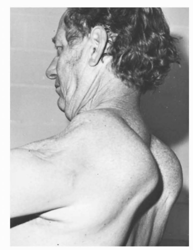
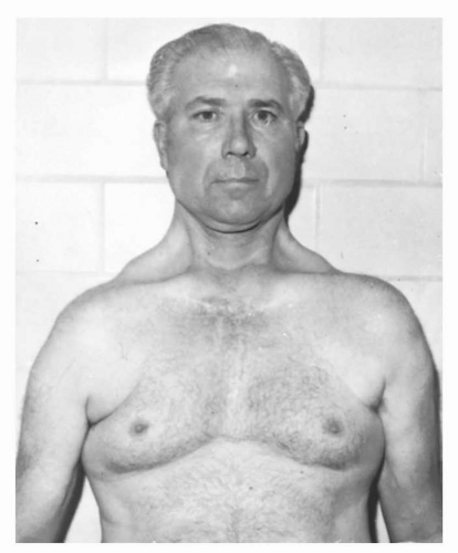
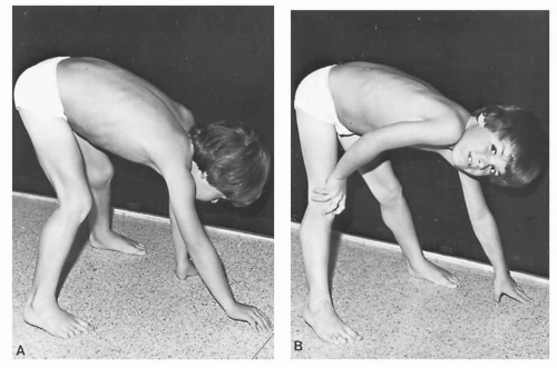
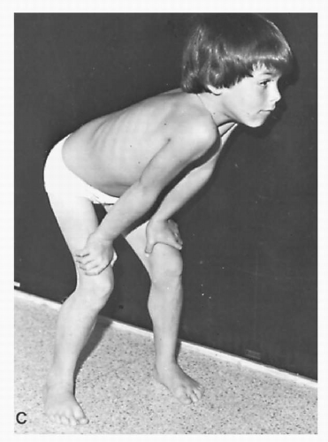
Proximal weakness is characteristic of many muscle disorders, the spinal muscular atrophies, and MG. These patients report difficulty in climbing stairs or arising from low chairs. When arising from a chair, they will lean forward and “push off” with their hands on the armrests. In arising from the floor or a squatting position, they may require one or more supports with the hands on the floor, knees, and thighs (Gowers maneuver; Fig. 17.1). The gait of a person with proximal weakness has a waddling appearance because of hip girdle weakness. Knee extensor weakness may cause the leg to “give out.” Patients may lock their knees to compensate, gradually leading to hyperextension (back-kneeing), which in turn produces an exaggeration of the lumbar lordosis. Shoulder girdle weakness produces difficulty in elevating the arms and may be accompanied by scapular winging (Fig. 17.2). With the arms hanging at the sides, the scapulae may slide laterally to produce a curving inward of the shoulders with the backs of the hands facing forward and an associated oblique “axillary crease” (Fig. 17.3). The high-riding scapulae produce a conspicuous “trapezius hump”; the clavicles may slope downward and stand out prominently from the atrophic neck musculature (Fig. 17.3).


FIGURE 17.1 Gowers maneuver displayed in arising from the floor (Duchenne muscular dystrophy).


FIGURE 17.1 Continued


FIGURE 17.2 Winging of the scapulae when the arms are elevated (fascioscapulohumeral dystrophy [FSH]).


FIGURE 17.3 Shoulder girdle weakness with “trapezius hump,” “step-sign” with prominent downsloping clavicles, and an oblique anterior axillary crease (LGD).

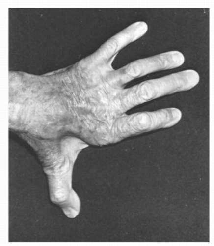
Distal weakness in the presence of atrophy is commonly seen in amyotrophic lateral sclerosis (ALS) (Fig. 17.4), inclusion body myositis (IBM), and myotonic dystrophy. Patients with ALS or IBM may present with unilateral symptoms but eventually develop weakness bilaterally. These patients find it difficult to manipulate small objects such as buttons or writing or eating utensils. They may complain of “dragging” their legs because of “foot drop” or of frequent tripping, particularly on uneven ground. With each step, the knees are raised high while the feet flap limply; shoe soles may show asymmetrical wear.

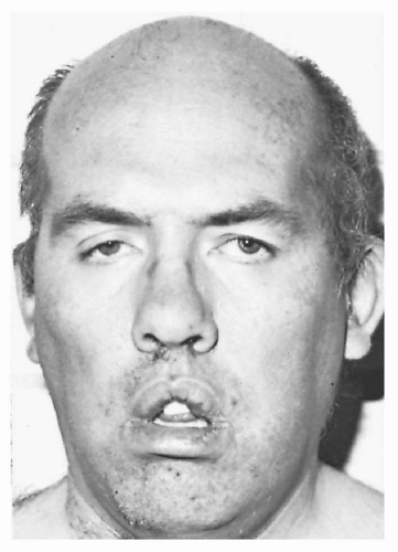
Cranial weakness may manifest as extraocular, facial, or oropharyngeal muscle weakness and is an important differential feature in diagnosis of various dystrophies (Figs. 17.5 and 17.6). Ptosis and ophthalmoparesis occur in several myopathic disorders and are very common in MG. Swallowing and speech changes can also be early signs of either ALS or MG.


FIGURE 17.4 Marked atrophy of the first dorsal interosseous muscle in motor neuron disease.


FIGURE 17.5 Typical facial appearance in myotonic dystrophy with frontal balding, temporalis and masseter atrophy, ptosis, and protuberant lower lip.


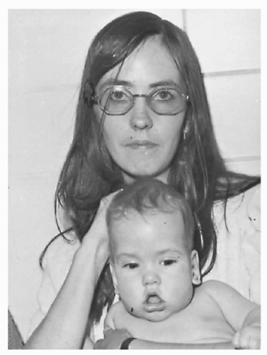
FIGURE 17.6 Myotonic dystrophy in mother (note “hatchet face”) and infant (note “shark mouth”). Diagnosis first made in the child suggested the same diagnosis in the mother.


Atrophy and Hypertrophy

The disuse of a limb will produce modest muscle atrophy, as is seen in nonneuromuscular conditions such as after casting of a bone fracture. The muscle retains much of its strength in disuse atrophy, and the apparently atrophied limbs of an elderly person may be surprisingly strong. In contrast, the striking muscular atrophy in patients with neuromuscular disease is associated with obvious weakness.

Atrophy of the muscles around the shoulder girdle commonly reveals underlying bony prominences. Flattening of the thenar eminence and guttering of the interossei can produce a wasted, clawlike deformity of the hand (Fig. 17.4). Several disorders produce characteristic appearances. Examples include “Popeye arms” (very atrophied biceps/triceps with a normal-appearing forearm) in facioscapulohumeral dystrophy, the “hatchet face” (temporalis wasting) appearance in myotonic dystrophy (Figs. 17.5 and 17.6), and
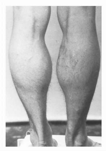
the enlarged calves of Duchenne dystrophy (DUD; Fig. 17.7). Diffuse hypertrophy of all limb muscles is commonly seen in myotonia congenita, rarely in hypothyroidism, or very rarely in amyloidosis.


FIGURE 17.7 Pseudohypertrophy of the calves (Duchenne dystrophy).


Pain, Stiffness, and Cramps

Pain is a nonspecific symptom that may be seen in many neuromuscular conditions. Inflammatory myopathies and other collagen vascular diseases may produce muscle pain and tenderness, but the absence of these symptoms does not exclude the diagnosis. Some patients may have difficulty distinguishing between pain and weakness because pain limits their ability to perform motor tasks. This may result in perceived weakness. Such individuals often prove to have normal strength or a “give-away” pattern of weakness, which suggests lack of full voluntary effort.

▪ SPECIAL CLINICAL POINT: Most patients with limb aching and pain without objective weakness or abnormally elevated muscle enzymes do not have a neuromuscular disease, and other possibilities should be considered, such as joint or soft tissue pathology.

In older patients, pain, aching, and stiffness in the shoulder and hip girdle muscles should suggest polymyalgia rheumatica, and this disorder is associated with a very high sedimentation rate. It is important to recognize because of its very specific treatment and rapid response to steroid medications. The diagnosis of fibromyalgia often is evoked, particularly in otherwise healthy individuals who have diffuse aches and pains without objective signs of weakness; however, the pathologic foundation of this entity remains controversial.

Stiffness may be a nonspecific symptom, or it may be a symptom of myotonia, a phenomenon consisting of a delayed relaxation of the muscle following voluntary contraction or percussion. This produces difficulty in releasing the grip or initiating movements after a period of rest and may point to a myotonic dystrophy or myotonia congenita.

Muscle cramp, a prolonged involuntary contraction, is a universal and generally benign symptom that occurs with increased frequency during unaccustomed exercise, “body building,” pregnancy, or electrolyte disturbance. It also may occur in hypothyroidism, partial denervation due to ALS or other less sinister types of nerve injuries, tetany (with hypocalcemia, hypomagnesemia, or alkalosis), and certain metabolic myopathies.


Muscle Twitching

Fasciculations are contractions of muscle fibers in a single motor unit. Fasciculations appearing in a strong muscle are usually benign and are exacerbated by many factors, including fatigue and caffeine intake. When they occur in a weak muscle, they may be associated with ALS, but they also may occur in the setting of root compression or peripheral nerve injury.


Infantile Hypotonia

The most frequent abnormality causing infantile hypotonia or a “floppy baby” is CNS disease, such as seen after perinatal asphyxia. The hypotonic infant may exhibit normal muscle strength, with the ability to lift its head or limbs against gravity. In the absence of other abnormalities, the prognosis for normal development may be excellent. In infants with obvious weakness, the underlying disorder may be spinal muscle atrophy or one of the congenital myopathies. Weakness of sucking and respiration are serious concomitant findings, which are usually fatal if undiagnosed and untreated.


Deformities

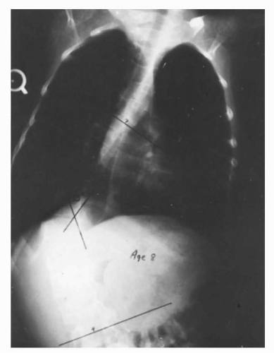
Neuromuscular disorders often are associated with skeletal deformities and should be suspected in a patient with unexplained hip dislocation, scoliosis (Fig. 17.8), contracture, or malformation of the feet (e.g., clubfoot, equinovarus, or pes cavus deformity). Arthrogryposis or multiple congenital limb deformities may occur with various diseases affecting any part of the motor unit.


FIGURE 17.8 Scoliosis in chronic infantile spinal muscular atrophy. The angle of curvature is measured and followed closely (55 degrees in this patient).

Only gold members can continue reading. Log In or Register to continue

Stay updated, free articles. Join our Telegram channel

Sep 8, 2016 | Posted by in NEUROLOGY | Comments Off on Neuromuscular Diseases

Full access? Get Clinical Tree

Get Clinical Tree app for offline access