Neurophysiological investigations
The electroencephalogram
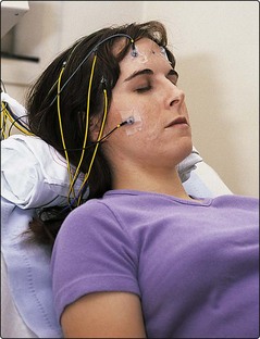
Method
The patient rests back and 20 electrodes are attached over the scalp with glue (Fig. 1). These are connected to a multichannel recorder, which generates a paper tracing or a computer record. This is often synchronized with a video recording of the patient. A skilled technician monitors the recording throughout, to detect and eliminate artefacts. The EEG is recorded with the patient’s eyes open and closed and several methods may be used to enhance sensitivity of the technique, routinely including forced hyperventilation for 3 min and stroboscopic photic stimulation at 1–50 Hz. Another technique to increase sensitivity is to deprive the patient of sleep before the EEG, then allow the patient to fall asleep during the recording.
EEG interpretation
Paroxysmal interictal EEG changes
Spikes and sharp waves may be focal (affecting only part of the brain) or generalized (simultaneously affecting all parts of the brain) (Figs 2 and 3). Focal spikes suggest epilepsy due to a focal disturbance and imply a focal structural cause; neuroimaging should be considered. Generalized spikes are seen as part of the generalized epilepsies, which usually start in childhood or adolescence (pp. 74–75
Stay updated, free articles. Join our Telegram channel

Full access? Get Clinical Tree


